
Rust in the Roots: The Copper-Iron Story Behind Hair Loss
31 October 2026
By Libbie Wallace
Hair loss can be a whole-body problem that shows at the scalp
Hair follicles contain some of the fastest-dividing cells in the body, and they depend on a steady supply of iron. Most of that supply doesn't come from food. Each day the body needs around 20–25 mg of iron, mainly to make new red blood cells, but absorbs only 1–2 mg from the diet. The rest is recycled.
That recycling happens in the reticuloendothelial system. Macrophages in the spleen, liver and bone marrow break down old red blood cells and release their iron back into the blood. Releasing it depends on copper: ceruloplasmin, a copper enzyme, converts the iron into a form its carrier, transferrin, can pick up as it leaves the macrophage. This step also depends on retinol. Retinol drives ceruloplasmin production, appears to be needed to make transferrin itself, and helps release recycled iron onto it. When copper or retinol runs short, often from high-dose zinc, gut problems, poor intake or too much iron from fortified foods and supplements, which itself lowers copper, iron stays locked inside the recycling cells and builds up in organs. The blood, and the follicles it feeds, run short. Poorly bound iron left behind in tissues then fuels free radicals, inflammation and scarring.
Routine testing isn't designed to see this. It measures what's in the blood, not what's sitting in the tissues. Serum ferritin, the usual hair loss test, is largely leaked from stressed and damaged cells. It isn't a store of iron, so no result can be taken as reassurance on its own. Copper, ceruloplasmin, zinc, RBC magnesium and retinol are rarely checked at all.
This post follows the copper-iron thread through the research. It covers how copper unlocks iron, a copper enzyme inside the hair follicle itself, what drives the scarring that makes pattern loss so hard to reverse, and what testing would actually reveal. Along the way, I'll be clear about what is proven, what is plausible and what is still waiting to be tested.
Copper keeps the iron recycling loop turning
Iron isn't meant to sit still. The body has no way to excrete it, so it reuses the same iron again and again. Old red blood cells are broken down by macrophages in the spleen, liver and bone marrow, and their iron is handed back to the bone marrow to build new ones.
The whole system is built for flow.
Ferritin is a holding bay, not a warehouse
Ferritin is usually called an iron "storage" protein. In the recycling system, it behaves more like a holding bay. When a macrophage breaks down an old red cell, it parks the iron in ferritin to keep it safe. A cargo protein called NCOA4 then delivers that ferritin to the cell's own recycling machinery. The cage is broken open and the iron is freed for export. This process is called ferritinophagy.
When this hand-over fails, the holding bay becomes a trap. Mice lacking NCOA4 build up ferritin and iron in several organs, especially in spleen macrophages, and become anaemic. Researchers showed the anaemia came from macrophages failing to release iron, not from a problem in the bone marrow. Inflammation does something similar. In lab studies, hepcidin switched off NCOA4 in macrophages, and ferritin piled up inside them.
Copper opens the exit door
Once freed, iron leaves the cell through a channel called ferroportin as ferrous iron (Fe2+). It must then be converted to ferric iron (Fe3+) before transferrin, the blood's iron carrier, can pick it up. That conversion is done by copper-containing enzymes called ferroxidases:
- Ceruloplasmin, made in the liver. It carries most of the copper in your blood.
- GPI-ceruloplasmin, a form anchored to the membranes of macrophages and liver cells, right where recycled iron leaves.
- Hephaestin, in the gut wall, which handles the small amount of iron absorbed from food.
Remove copper and these enzymes lose function. Copper-deficient rats develop low blood iron and anaemia while iron builds up in the liver. Ferroportin itself also appears less stable when ferroxidase activity drops, which traps even more iron.
The clearest human proof is a rare genetic condition, aceruloplasminemia, where ceruloplasmin is missing entirely. People with it have low blood iron and low transferrin saturation, yet iron steadily accumulates in the liver, pancreas and brain.
Retinol helps release recycled iron
Retinol is a less-known partner in this loop. It matters at two points: making transferrin, the carrier, and getting recycled iron out of storage and onto it.
Researchers reviewing the retinol-iron link proposed that a lack of retinol may directly impair transferrin production, and that restoring retinol may raise plasma transferrin. In cell studies, retinol stimulated transferrin production. Transferrin and the retinol carrier protein also fall together during infection. The practical point: a low transferrin, or a low total iron-binding capacity, shouldn't be put down to protein intake or inflammation alone. Retinol deficiency is worth ruling out.
In rats, retinol deficiency disrupted the signals that control hepcidin, and iron was held back in the spleen while the rest of the body ran short. Retinoic acid also stimulates ceruloplasmin production, linking retinol directly to copper's role at the exit door.
Human trials support this:
- In Moroccan children low in both retinol and iron, retinol supplementation raised haemoglobin and lowered serum ferritin. Researchers read this as iron being mobilised from liver stores, without any change in total body iron.
- In a randomised trial of 250 Malaysian schoolchildren, one high dose of retinol raised serum iron and transferrin saturation. Iron deficiency anaemia fell by about 22%, against 2.3% in the placebo group.
- A 2019 systematic review concluded that retinol supplementation alone may reduce anaemia in people with low serum retinol.
In other words, some "iron deficiency" may really be iron that can't get out of storage, because retinol, copper or both are missing.
Three places the loop can jam
- Ferritin isn't broken down, so iron stays parked in the holding bay.
- Ferroportin is destroyed by hepcidin during inflammation, so the exit door closes.
- Iron can't be converted on its way out, because copper and ceruloplasmin are lacking.
Each one leaves iron stuck in tissues while the blood, and the hair follicles, run short. That's why taking more iron so often fails, and can add to the build-up. Researchers note that the anaemia of copper deficiency doesn't improve with iron replacement. The problem is iron use and recycling, not iron supply.
The hair follicle has its own copper-iron enzyme
The most striking evidence for this whole idea rarely appears in hair loss articles. In 2019, researchers described a boy with sparse, coarse, twisted and breaking hair. They traced it to faults in a gene called HEPHL1, a newly characterised copper-containing ferroxidase from the same family as hephaestin.
What they found matters:
His skin cells built up iron inside them, exactly what you'd expect when a ferroxidase stops working.
Those same cells showed reduced activity of lysyl oxidase, another copper-dependent enzyme.
- Mice bred without the gene were healthy but grew curly whiskers, echoing the boy's hair.
The authors concluded that ferroxidase activity plays a role in hair growth itself, and that HEPHL1 may have a specific job in the follicle that other enzymes can't cover. In plain terms: the follicle appears to need its own copper-powered way of handling iron.
This fits an older clue. Menkes disease, a genetic disorder of copper transport, causes brittle, twisted, depigmented "kinky" hair. The copper link was spotted when researchers noticed copper-deficient sheep grew similarly kinked wool. Several copper enzymes help explain the hair changes:
| Copper enzyme | Its job | What failure looks like in hair |
|---|---|---|
| Lysyl oxidase | Cross-links collagen and structural proteins | Fragile, twisted shafts |
| Tyrosinase | Makes melanin | Loss of hair colour |
| Cytochrome c oxidase | Final step of mitochondrial energy production | Less energy for fast-dividing follicle cells |
| Superoxide dismutase (Cu/Zn) | Neutralises free radicals | Less protection from oxidative stress |
A word of proportion: Menkes and HEPHL1 variants are rare genetic conditions, and they mostly affect hair structure rather than causing ordinary thinning. They don't prove that mild copper shortfalls cause everyday hair loss. What they do show is that the follicle depends on copper, and that copper and iron handling are tied together right at the root.
How copper quietly runs low
A plain dietary copper shortage is uncommon. Most documented cases come from something blocking absorption, and the biggest culprit is zinc.
Zinc switches on a protein in gut cells called metallothionein. Metallothionein binds copper even more tightly than zinc, so the copper gets locked inside the gut lining and is shed when those cells turn over. High zinc intake can therefore create a copper deficiency on its own.
The irony for hair is obvious. Zinc is one of the most popular hair loss supplements. Taken at high doses for months, it may work against the very system it's meant to support.
Excess iron drains copper too. In growing rats and mice, high-iron diets reduced copper absorption, lowered ceruloplasmin activity and caused copper-deficiency anaemia. Disturbances appeared with iron at only 9.5 times the requirement. Giving copper prevented the damage, showing that copper loss was the cause. The researchers flagged iron-fortified refined grains and the iron in daily multivitamins as a real-world concern.
Human studies are less consistent. In breastfed infants, iron supplements reduced the activity of copper-dependent superoxide dismutase, while another study found no change in copper absorption. A 2026 review concluded that people whose copper is already marginal, or who take high-dose iron for long periods, are the most vulnerable. That's exactly the group this blog is about.
The problem with iron-fortified food
In the UK, iron is added by law to all non-wholemeal wheat flour. That means white and brown bread, cakes, biscuits, pastry, pizza and most processed foods made with flour. The current legal minimum is 1.65 mg of iron per 100 g of flour. From 13 December 2026, it rises to at least 2.10 mg, an increase of more than a quarter. Many breakfast cereals add iron on top of that. Someone eating a typical diet can be taking in added iron at almost every meal without realising it.
This isn't the iron found naturally in food. The permitted forms are ferric ammonium citrate, ferrous sulphate and iron powder. These are the same kinds of iron used in tablets, without the proteins and cofactors that come with iron in whole foods.
For the copper-iron loop, fortified food is a problem in four ways:
- It drains copper. In animal studies, excess iron reduced copper absorption and ceruloplasmin activity at intakes not far above requirements.
- It loads a jammed system. When copper or retinol is low, recycled iron already can't leave the tissues. Every extra dose of incoming iron adds to the build-up.
- It feeds free radicals. Iron that isn't properly bound is exactly the form Douglas Kell links to hydroxyl radical damage and chronic inflammation.
- It's invisible. Iron is listed among the ingredients, but almost nobody counts how much they eat. It's rarely considered when someone presents with hair loss.
Practical steps:
- Choose wholemeal flour and wholemeal bread, which are exempt from mandatory fortification.
- Check breakfast cereal labels for added iron, and pick unfortified options.
Base meals on whole foods rather than processed, flour-based products.
Don't reach for iron when blood iron is low. Low blood iron is often a recycling problem, not a shortage: check retinol first, then copper, ceruloplasmin and CRP. The one thing to rule out alongside is blood loss, such as heavy periods or bleeding from the gut.
Denture adhesive is the best-documented example of how far this can go. Case reports describe people who used zinc-containing creams heavily for years and developed copper deficiency with anaemia, low white cell counts and nerve damage. In one UK case, a man had used two to four tubes a week for 15 years and ended up using a wheelchair. Another woman's anaemia kept worsening even while she took iron, until her copper deficiency was found.
Other routes to low copper include: - Gastric bypass and other stomach or bowel surgery - Coeliac disease and other malabsorption - Long-term high-dose zinc from supplements, cold remedies or stacked multivitamins
Note what those case reports share: patients were often treated with iron or B12 first, and it didn't help. Copper deficiency is easy to miss because its blood picture can mimic other deficiencies.
Where this meets pattern hair loss
Androgenetic (pattern) hair loss is driven by DHT shrinking genetically sensitive follicles. The copper-iron idea doesn't replace that. The question is whether a copper-iron problem could add to it.
The human data so far is thin and mixed: - A 2024 Pakistani study comparing several types of alopecia with healthy controls found significantly low serum copper only in the male pattern hair loss group. Its authors suggested men with pattern loss should have copper checked, and supplement only if it's low. - A 2024 meta-analysis of 34 studies (4,931 people) on alopecia areata found lower zinc and vitamin D in patients, but no consistent difference in copper. That was autoimmune hair loss, not pattern loss, so it doesn't settle the question either way.
The proposed mechanism goes like this. Copper shortfall weakens copper-zinc superoxide dismutase, an antioxidant, while iron left loose inside cells can generate free radicals through the Fenton reaction. Follicles already stressed by DHT would then face extra oxidative damage and lower energy output.
It's a coherent hypothesis, and each piece is real biology. But no study has yet measured iron build-up in the scalps of people with pattern hair loss, or shown that correcting copper slows it. Scarring around follicles (perifollicular fibrosis) is real and well documented in pattern hair loss, as the fibrosis section below explains. What hasn't been shown is that iron drives it. Treat this as a promising line of inquiry, not a settled cause.
A practical clue worth watching: pattern loss affects the top and front, while shedding from a nutritional or stress cause tends to be diffuse, including the back and sides. Seeing both at once suggests more than one process is at work.
Endothelin-1: timekeeper, wake-up call and scar-maker
Endothelin-1 (ET-1) has at least three jobs that matter for hair, and they pull in different directions. That's why simple "ET-1 kills follicles" claims don't hold up, and why the real story is more interesting.
Job 1: ending the growth phase on schedule
A 2023 Nature Cell Biology study from Mount Sinai showed how a follicle shrinks back at the end of each growth phase. The dermal sheath, a thin sleeve of smooth muscle around the follicle, contracts and pushes the follicle upwards. The trigger is ET-1, released by progenitor cells at the narrowing "bottleneck" of the regressing follicle.
The evidence is strong:
- Both endothelin receptors (ETA and ETB) are concentrated in dermal sheath cells.
- ET-1 contracted these cells in a dose-dependent way in the lab.
- Blocking both receptors, with drugs or by deleting the genes, stalled regression. Knocking out just one receptor didn't, because each covers for the other.
- When receptor blockers were applied to mouse skin, many follicles were still stuck mid-regression when the next resting phase began.
This matches decades-old patent work in guinea pigs, where injected endothelin pushed growing follicles into rest.
Job 2: waking resting follicles up
A 2024 FASEB Journal study found the opposite effect from a different source. ET-1 was produced more by epidermis over actively growing follicles. It made dermal papilla cells multiply and switched resting follicles into a new growth phase in mice, acting through the PI3K/AKT pathway. The authors suggested ET-1 could even be a hair-growth treatment target.
So the same molecule appears to close one cycle and open the next. Where it comes from and when it arrives seem to decide which.
Job 3: driving fibrosis
This is the part almost no hair article mentions. Outside the hair world, ET-1 is one of three main drivers of skin fibrosis, alongside TGF-β and connective tissue growth factor (CTGF). Research in scleroderma, the classic fibrotic skin disease, shows:
- ET-1 makes skin fibroblasts produce more collagen, fibronectin and fibrillin.
- TGF-β switches on ET-1 production in skin fibroblasts, and part of TGF-β's scarring effect runs through that ET-1.
- The two receptors physically cooperate to turn quiet fibroblasts into collagen-producing myofibroblasts.
- The endothelin blocker bosentan prevented skin fibrosis in mice and reversed the scarring profile of patients' fibroblasts in the lab.
What this means for hair
Put together, a plausible picture emerges, though it hasn't been tested directly in the scalp. In a balding scalp, TGF-β is raised (see the next section). TGF-β is known to raise ET-1 in skin fibroblasts. Chronically raised ET-1 could then do two harmful things: keep triggering early regression, and push collagen build-up around the follicle.
The honest gap: no published study has yet measured ET-1 levels in balding versus non-balding human scalp. If that comparison is ever made, it could explain how hormones, scarring and shortened growth cycles connect. For now, it's a hypothesis worth watching, not a basis for treatment. Endothelin-blocking drugs are prescription medicines for lung blood pressure with serious side effects, and they are not hair treatments.
What really causes scalp fibrosis
Scarring around the follicle is a real, measured feature of pattern hair loss. It isn't just an internet theory. Biopsies show a thickened dermal sheath made of extra collagen, called perifollicular fibrosis. It matters because it predicts how well treatment works.
In studies by Whiting, around 40% of pattern hair loss biopsies showed inflammation around follicles. Men with that inflammation and fibrosis responded less well to minoxidil: 55% regrew hair, against 77% of those without it. An Egyptian study of 40 men found inflammation was almost always present in early cases and was linked to later fibrosis. In advanced cases, the collagen sheath thickened further and sometimes destroyed the follicle entirely.
The research points to six overlapping drivers. They're listed from strongest evidence to weakest.
1. Androgens switch on TGF-β (strong evidence)
TGF-β is the body's master scar signal. A 2002 study showed androgens make dermal papilla cells from balding scalp release TGF-β1, which suppresses the growth of surrounding hair cells. Blocking TGF-β1 with an antibody reversed that suppression. Androgens also drive TGF-β2, DKK-1 and IL-6 from balding papilla cells, all of which inhibit growth.
In 2006, Korean researchers showed testosterone raised TGF-β1 by 82% in scalp fibroblasts and boosted type I collagen production. Finasteride pretreatment cut the TGF-β1 rise by about 30%. That suggests part of finasteride's benefit may come from slowing fibrosis, not only from lowering DHT.
2. Oxidative stress feeds TGF-β (strong lab evidence)
This is where the copper-iron story connects. A 2013 study found androgens raise reactive oxygen species (free radicals) in dermal papilla cells. Those free radicals, in turn, drive TGF-β1 release. Antioxidant treatment reduced it.
A 2022 study found balding dermal papilla cells were under significantly higher oxidative stress than non-balding cells from the same men. Their mitochondria were also struggling. Activity of energy complexes I, IV and V was reduced, and so was ATP output. Complex IV is cytochrome c oxidase, the copper-dependent enzyme discussed earlier. Antioxidants improved how these cells took up fuel.
No one has yet tested whether copper status affects complex IV in balding scalp. But the overlap is striking: a weaker copper-dependent enzyme sits in exactly the cells that are failing.
3. Low-grade inflammation (good evidence)
Low-grade inflammation around the upper follicle, with T cells and macrophages, appears early in pattern loss. Over time, as fibrosis increases, the inflammation tends to fade, as if it leaves the scar behind.
A 2012 University of Pennsylvania study found prostaglandin D2 (PGD2), an inflammatory lipid, was three times higher in bald scalp than haired scalp in the same men. PGD2 inhibited hair growth in human follicles and in mice, through a receptor called GPR44. Mice engineered to overproduce it in skin went bald. The authors noted this is still a correlation in humans.
4. Shrinking blood supply (good evidence, direction debated)
Balding scalp has measurably less blood flow and oxygen. A Danish study found subcutaneous blood flow was much lower in early male pattern baldness, and later work found lower oxygen levels in balding areas. A 2022 study showed how this may start: androgen-activated papilla cells send TGF-β signals that kill the blood vessel cells next to them. The vessels regress at the very start of miniaturisation.
The debate is over cause and effect. A smaller follicle needs less blood, so reduced flow could be a result. But the 2022 vessel findings suggest vascular loss happens early, not only at the end.
5. Mechanical tension from the scalp (hypothesis, modelling only)
Pattern hair loss only occurs over the galea aponeurotica, the tough sheet of tissue that stretches between the forehead and back-of-head muscles. A 2015 engineering study modelled the stress on that sheet and found it matched the Norwood balding pattern closely. The proposed link is a protein called Hic-5, which raises follicles' sensitivity to androgens and can be activated by stretching. Hic-5 is higher in papilla cells from bald frontal scalp than from the back of the head.
This is a computer model plus biological plausibility. It hasn't been tested by measuring tension in real scalps, and it doesn't explain why women's patterns differ. A 10-man pilot study of scalp Botox, which relaxes these muscles, reported some benefit, but larger trials are needed.
6. Tissue iron (hypothesis, borrowed from other tissues)
This is the link the copper-iron idea predicts, and there's real evidence for iron causing fibrosis elsewhere in the skin. In chronic venous disease of the legs, iron deposits are absent in mild skin changes. But they are always present in lipodermatosclerosis, the hard, scarred skin of advanced disease, and in ulcers. That scarred skin also shows raised TGF-β1 and collagen. Researchers have also found that iron creates oxidative stress in skin fibroblasts and pushes cells towards a scar-forming state. They propose tissue iron as a missing link between small-vessel damage and fibrosis in scleroderma.
The mechanism is the same one described above: iron generates free radicals, free radicals activate TGF-β, and TGF-β builds collagen. What's missing is any measurement of iron in balding scalp. A simple iron stain on scalp biopsies could test this. Until someone does it, iron remains a plausible contributor, not a proven one.
Five drivers converge on one scar signal, TGF-β1
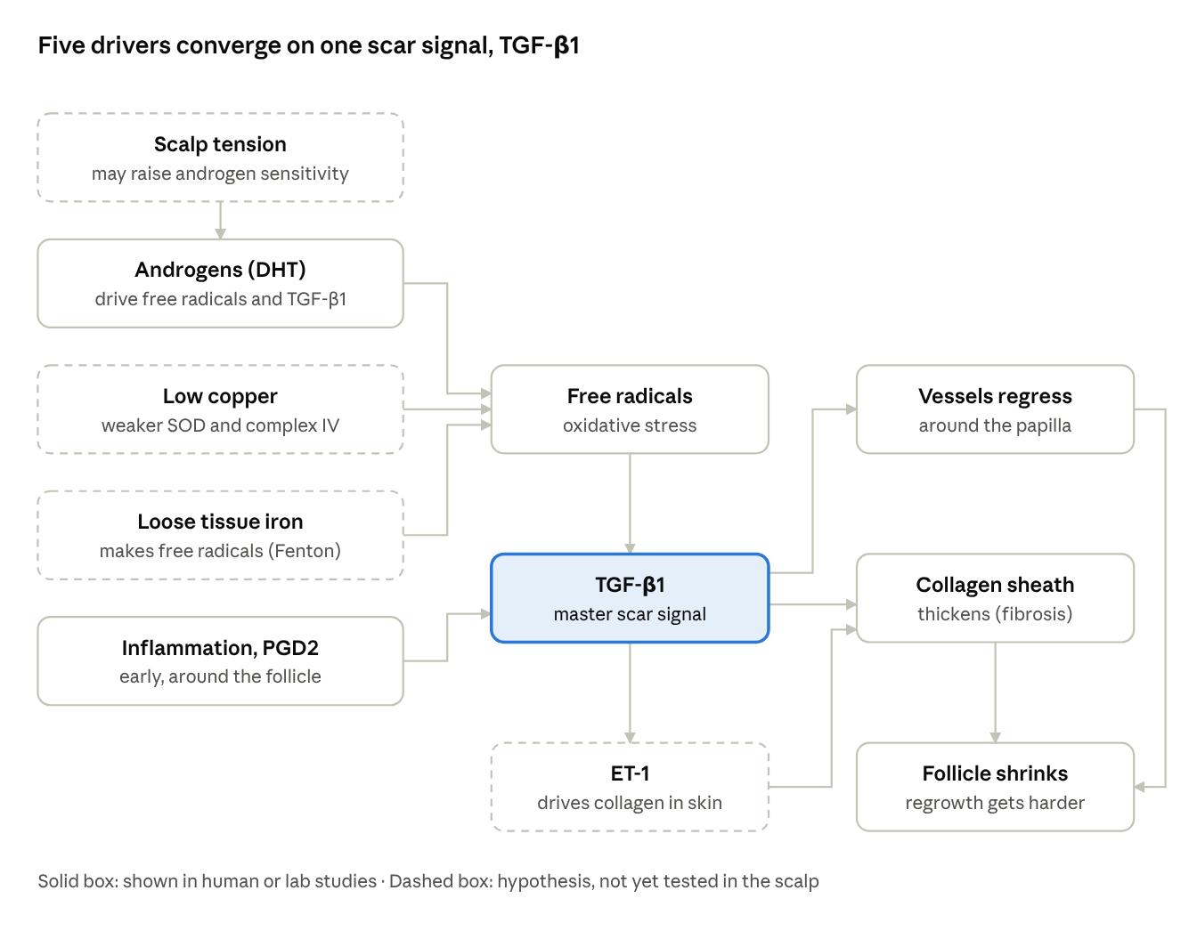
Solid box: shown in human or lab studies · Dashed box: hypothesis, not yet tested in the scalp
scalp fibrosis drivers · 5 inputs, 1 hub
Most routes run through free radicals and TGF-β1. Reduced blood supply appears here as a result of TGF-β1 rather than an input, and copper is shown separately from iron. The dashed boxes, including copper, iron and ET-1, are the links still waiting for scalp studies.
Other triggers hiding in plain sight
Before chasing minerals, it's worth ruling out the two most common triggers of sudden diffuse shedding: medications and stress. Both typically show up two to three months after the trigger, which is why people rarely connect them.
Medications most often linked to shedding include:
- Heart and blood pressure drugs: beta-blockers (propranolol, atenolol, metoprolol), ACE inhibitors, warfarin and heparin
- Hormonal treatments: some contraceptive pills (and stopping them), testosterone and anabolic steroids, HRT
Mental health and neurology drugs: SSRIs such as sertraline and fluoxetine, lithium, sodium valproate, carbamazepine
Retinoids such as isotretinoin, and high-dose retinol supplements
- Thyroid treatment, alongside the thyroid condition itself
Drug-related shedding is usually reversible once the cause is changed, but never stop a prescribed medicine without talking to your prescriber.
Stress can push large numbers of follicles out of the growth phase at once (telogen effluvium). Daily shedding can rise from a typical 50-100 hairs to several hundred. Severe stress is also a recognised trigger for alopecia areata. Recovery usually starts within months of the stressor easing, though full density can take six to twelve months to return.
These triggers also stack. Someone on long-term medication, under strain, and taking high-dose zinc "for their hair" may be dealing with three overlapping problems at once.
What the botanicals can and can't do
Plant-based options have more real data behind them than sceptics assume, and less than sellers claim. Here's where the main ones stand.
| Botanical | How it's used | Best human evidence | Strength of evidence |
|---|---|---|---|
| Pumpkin seed oil | Oral capsules | 2014 double-blind trial, 76 men: 400 mg daily for 24 weeks raised hair counts 40% vs 10% on placebo | Promising, one main trial |
| Saw palmetto | Oral or topical | 2020 systematic review: 5 small trials and 2 cohort studies reported benefit, mostly in combination products | Limited, mostly subjective measures |
| Rosemary oil | Topical | 2015 trial, 100 men: similar hair count gains to 2% minoxidil at 6 months, with less itching | Promising, no placebo arm |
| Ginger, tea tree, aloe | Topical tonic | No direct hair-growth trials in people | Soothing and anti-dandruff, growth unproven |
A few points the marketing tends to skip:
- Pumpkin seed oil and saw palmetto are thought to work by inhibiting 5-alpha reductase, the enzyme that makes DHT. They target pattern loss, not shedding from nutrient or stress causes.
- The pumpkin seed oil trial used a combination product that contained other ingredients, so the effect can't be credited to pumpkin seed alone.
- Rosemary was compared with 2% minoxidil, not the stronger 5%. Mouse studies suggest it also inhibits 5-alpha reductase.
- Taking an oil orally rather than applying it changes how it works. An oral DHT-blocker acts body-wide, so it's worth mentioning to anyone prescribing for you, especially before PSA testing.
- None of these will correct a copper or iron problem. At best, they address the DHT side while the internal picture is sorted separately.
A sensible combination, then, is an oral DHT-modulating supplement plus a light topical tonic, used consistently for at least six months before judging results.
Tests worth asking about
A basic full blood count and ferritin won't reveal a copper-iron transport problem. If the picture doesn't add up, these are the markers to discuss with your clinician:
| Marker | Why it matters here |
|---|---|
| Serum copper | Direct measure of circulating copper |
| Ceruloplasmin | Falls in copper deficiency; the key ferroxidase that moves iron out of tissues |
| Serum zinc | High zinc points to zinc-induced copper loss |
| Ferritin | Iron stores, but also an acute-phase protein; never read it alone |
| CRP (and ESR) | Shows whether inflammation is pushing ferritin up or trapping iron via hepcidin |
| Serum iron, TIBC, transferrin saturation | How much iron is actually circulating and available |
| RBC magnesium | Intracellular magnesium; deficiency linked to iron build-up in animal studies |
| Retinol | Needed for transferrin and ceruloplasmin production; check it whenever transferrin or TIBC is low |
| Marker | Why it matters here |
|---|---|
| Full blood count with white cells | Copper deficiency can lower white cells as well as red |
| Thyroid panel (TSH, free T4, free T3) | Thyroid problems mimic nutritional shedding |
| Free and total testosterone, SHBG, DHEA-S | The hormonal side of pattern loss, especially in women |
The pattern to look for: normal or high ferritin alongside low transferrin saturation, low copper or ceruloplasmin, and high zinc. That combination suggests iron is stored but not moving. Always check CRP at the same time, so inflammation isn't mistaken for healthy iron stores.
Practical tips for accurate results:
- Stop biotin for a few days beforehand. It interferes with several hormone and thyroid assays.
- Book a morning appointment, as iron and testosterone vary through the day.
- Tell the lab or clinician about every supplement you take, including zinc lozenges and denture creams.
If copper does come back low, correct it with professional guidance rather than self-dosing. Excess copper is harmful too, and the underlying cause, such as zinc, needs addressing first.
What the studies didn't measure
The research linking iron to hair loss rests almost entirely on one blood test: serum ferritin. A 2003 study judged iron stores in women with hair loss by ferritin alone, and a 2025 study measured only ferritin and haemoglobin. None of the studies I found measured ceruloplasmin, red cell magnesium or retinol. None measured iron in organs or in the scalp.
The one study that did look wider is telling. In women with chronic shedding, a Turkish study measured copper as well as iron and found low zinc, low copper and low ferritin. Copper was low in the same women whose low ferritin usually gets the blame.
Ferritin means little without inflammatory markers
Ferritin is an acute-phase protein. It rises with inflammation, infection, liver stress and metabolic problems, regardless of how much usable iron you have. That's why it should always be read alongside CRP, and ideally ESR. Read alone, it misleads in both directions:
- A "normal" ferritin with a raised CRP can hide depleted stores, because inflammation pushes the number up.
- A high ferritin with low transferrin saturation shows iron that is stored but not moving. It can come from inflammation, from ceruloplasmin failure, or both.
The second pattern deserves more attention than it gets. Inflammation raises a liver hormone called hepcidin, which destroys ferroportin, the channel iron uses to leave cells. Ceruloplasmin is needed to keep ferroportin stable at the cell surface. So inflammation and copper shortfall are two separate routes to the same result: iron locked inside tissues while the blood runs short. Standard reporting often treats the two as unrelated.
Serum ferritin isn't stored iron
Inside cells, ferritin is a protein cage that safely stores up to thousands of iron atoms. The ferritin measured in blood is different. It is almost entirely iron-poor and plays no part in moving iron around the body. Ferritin isn't made in the blood at all.
In 2014, researchers at the University of Manchester and the University of Pretoria argued that serum ferritin is mainly a leakage product from damaged and stressed cells. It has dumped most of its iron on the way out. On this view, a raised serum ferritin is a sign of cell damage, and potentially of loose, reactive iron released inside tissues.
This matters most when ferritin is raised but CRP is normal. CRP picks up acute inflammation. It can miss slower damage from metabolic strain or iron loading in organs. In a study of fatty liver disease, serum ferritin had no relationship with CRP, ESR or the degree of liver inflammation. Instead, it tracked the iron actually stored in the liver. A raised ferritin with a normal CRP shouldn't be shrugged off. It's a reason to look for organ dysregulation, including iron accumulation, and to check ceruloplasmin and copper.
Ferritin is also an unreliable guide to how much iron is stored. One documented patient had a normal ferritin despite iron stores six to twelve times higher than normal. Ferritin only fell once those stores were depleted by repeated venesection. The test tracks depletion better than it tracks overload. The practical rule: no ferritin result, high, "normal" or low, is reassurance on its own. Every result needs reading against CRP, transferrin saturation, copper and ceruloplasmin before anyone can say iron is being handled well.
The cofactors nobody checks
Two more nutrients affect the copper-iron system. The evidence comes from controlled animal studies, the same foundation much of our understanding of copper and iron rests on. Human studies are the next step and haven't yet been done:
Retinol. Retinol-deficient rats stored more iron in the liver and spleen while serum iron fell. Retinoic acid also stimulates ceruloplasmin production in rats.
Magnesium. Magnesium-deficient rats developed fragile red cells that broke down early, with iron building up in the liver and spleen. Red cell (RBC) magnesium is often preferred to serum magnesium because most magnesium sits inside cells. But labs note it isn't yet a fully validated marker.
How organ iron can actually be assessed
Iron in organs can't be read from a blood test. It can be measured by liver MRI, which is how aceruloplasminemia is confirmed, or by iron staining of a tissue biopsy. Neither is routine, and no study has yet applied either to people with hair loss. Until someone does, "no iron accumulation" in hair loss is an assumption, not a finding.
The bigger picture: Douglas Kell's iron research
The scientist who has argued this case most consistently isn't a hair specialist. Douglas Kell is Research Professor in Bioanalytical Science at the University of Manchester. With physiologist Etheresia Pretorius, he has spent well over a decade arguing that iron in the wrong form is a common thread running through chronic disease.
"Iron behaving badly" (2009)
Kell's key insight is that the problem isn't iron itself. It's poorly liganded iron, iron that isn't properly bound to the proteins meant to hold it. Normal metabolism constantly produces peroxide and superoxide. On their own, these aren't very damaging. But when they meet loose iron, they produce the hydroxyl radical, one of the most destructive molecules in biology.
Kell reviewed evidence across cardiovascular, metabolic and neurological diseases, where iron is found at the sites of plaques and lesions. He argued that their shared features point to a common cause: iron-driven free radical production.
Two of his points matter directly for hair:
Antioxidants can backfire. Where loose iron is present, substances that are normally antioxidants can act as pro-oxidants. Reducing oxidative stress needs both antioxidants and properly bound iron. This may help explain why antioxidant supplements give such mixed results.
Damage can become self-sustaining. Free radicals and loose iron can feed each other in runaway cycles that are hard to stop unless several points are tackled at once.
Kell also noted that before publication, the paper was turned away by three journals without even being sent for external review. New systems-level ideas often struggle against established thinking.
Serum ferritin as a damage signal (2014)
As described above, Kell and Pretorius argued that serum ferritin is mostly leaked from damaged cells, having dumped most of its iron. That turns a routine blood test into a possible signal of tissue damage and iron release.
Iron dysregulation and dormant microbes (2018)
Their most ambitious idea is the Iron Dysregulation and Dormant Microbes (IDDM) hypothesis. It proposes that stress-induced iron dysregulation can reawaken dormant microbes the body has picked up through past infections or an unbalanced gut. Once active, these release potent inflammatory triggers, such as lipopolysaccharide (LPS). This leads to abnormal blood clotting, amyloid formation and cell death. They have reported LPS sitting alongside abnormal fibrin fibres in the blood of people with Parkinson's, Alzheimer's and type 2 diabetes.
What this means for hair
Kell hasn't studied hair loss directly. But his framework sharpens this blog's argument in three ways:
"Iron accumulation" becomes more precise. The danger is iron that isn't properly bound, not simply the total amount. Ceruloplasmin, and therefore copper, is part of how iron stays safely handled.
It explains the free-radical hub. The diagram above has free radicals at the centre of follicle fibrosis. Kell's chemistry explains why loose iron is such a powerful source of them.
It joins up inflammation and iron. Low-grade inflammation, gut health and iron handling become parts of one system, not separate problems.
His ideas remain a minority view, built mainly from wide-ranging literature reviews and laboratory work. Large clinical studies haven't tested them. They are, though, published in peer-reviewed journals, and they line up closely with the copper-iron pattern in this blog.
Connecting the dots
Step back from the individual studies and one theme runs through all of them: copper deficiency and iron accumulation. Hair loss is usually treated as a scalp problem with a hormone cause. This pattern suggests it can also be one visible sign of a system-wide problem.
The pattern
Copper-dependent enzymes keep turning up. Superoxide dismutase protects against free radicals. Complex IV makes energy and is underperforming in balding scalp cells. Lysyl oxidase builds hair structure. HEPHL1 handles iron inside the follicle. All of them need copper.
Iron trapping is well documented. When ceruloplasmin fails, iron builds up in liver, pancreas and brain while blood iron falls. Inflammation does the same through hepcidin.
Both feed the same damage. Loose iron and weak antioxidant defences both generate free radicals. Free radicals switch on TGF-β1, the scar signal at the centre of follicle fibrosis.
The effects are multi-system. In documented cases of zinc-induced copper deficiency, patients developed anaemia that didn't respond to iron, low white cell counts, nerve damage and cognitive problems. Hair is one tissue among many that depends on the same enzymes.
Where DHT fits
Pattern hair loss still requires DHT. Men castrated before puberty, and men born unable to make DHT, don't develop it. So the strongest version of this idea is that copper deficiency and iron accumulation act as an amplifier. They add oxidative stress, energy failure and scarring on top of the hormonal signal.
That would explain several things the hormone-only model struggles with: - Why some people lose hair far faster than family history predicts - Why many respond poorly to finasteride and minoxidil, especially once fibrosis has set in - Why diffuse shedding so often overlaps with pattern loss - Why iron tablets often fail to stop shedding, and why high-dose zinc can make things worse
Why this hasn't been tested
The tests that would reveal this pattern aren't routine. Copper, ceruloplasmin, zinc, RBC magnesium, retinol and inflammatory markers are rarely ordered together. Organ iron is almost never measured. A theory can't be ruled out by studies that never measured it.
Predictions that would prove or disprove it
A good hypothesis makes predictions. This one makes four:
- Iron staining of scalp biopsies should show more iron in balding scalp than at the back of the head.
- People with pattern hair loss should have lower ceruloplasmin and copper, or higher zinc, than matched controls, once ferritin is read alongside CRP.
- Balding dermal papilla cells should show reduced HEPHL1 and ferroportin activity.
- Correcting copper and its cofactors in people with confirmed low levels should slow thinning more than standard treatment alone.
Each of these is achievable with existing methods. Until they are done, copper deficiency and iron accumulation remain an unproven but coherent explanation, and one that conventional testing is not set up to find.
The bottom line
Copper is the overlooked partner in the iron story, and the hair follicle seems to rely on both. That much is grounded in solid biochemistry, rare genetic diseases and documented cases of zinc-induced copper loss.
What we still don't know: - Whether mild, everyday copper shortfalls contribute meaningfully to common hair loss - Whether iron actually builds up in the scalp in pattern hair loss - Whether correcting copper improves hair density in people without frank deficiency - How ET-1 signalling changes in thinning scalps, and whether minerals influence it
What you can act on now: 1. Check you aren't taking more zinc than you need, from any source. 2. If iron supplements haven't helped your shedding, ask about copper, ceruloplasmin and zinc. 3. Rule out medications, thyroid and recent stressors before blaming genetics. 4. If you try botanicals, choose the ones with trial data and give them six months.
The most useful shift is in the question itself. Instead of "do I have enough iron?", ask "can my body actually use the iron it has?"
This article is for education and exploration, not individual medical advice. Please discuss testing and supplements with a qualified clinician.
References
Broderius et al. — Perinatal copper deficiency, hypoferremia and hepcidin in rats
- Aceruloplasminemia: iron accumulation and chelation therapy
- Sharma et al. 2019 — Biallelic HEPHL1 variants impair ferroxidase activity and cause an abnormal hair phenotype
- Menkes kinky hair disease case report
- Hypocupraemia secondary to zinc-containing dental adhesive (BMJ Case Reports, 2021)
- Zinc-induced copper deficiency, sideroblastic anaemia and neutropenia
- Evaluation of copper and zinc in alopecia (JIIMC, 2024)
- Serum trace elements and alopecia areata: meta-analysis (J Cosmet Dermatol, 2024)
- Mount Sinai — Progenitor-derived endothelin controls dermal sheath contraction (Nature Cell Biology, 2023)
- Cho et al. 2014 — Pumpkin seed oil for androgenetic alopecia, randomised trial
- Evron et al. 2020 — Saw palmetto systematic review in alopecia
- Panahi et al. 2015 — Rosemary oil vs minoxidil 2%
Added for the endothelin-1 and fibrosis sections:
- Rendl lab, Mount Sinai — Progenitor-derived endothelin controls dermal sheath contraction (Nature Cell Biology, 2023)
- Gao et al. — Epidermal ET-1 activates resting hair follicles via PI3K/AKT (FASEB J, 2024)
- Akamata et al. — Bosentan reverses the pro-fibrotic phenotype of scleroderma dermal fibroblasts (2014)
- Endothelin-1, TGF-β and CTGF in connective tissue fibrosis (Arthritis Res Ther)
- Inui et al. — Androgen-inducible TGF-β1 from balding dermal papilla cells (FASEB J, 2002)
- Yoo et al. — Perifollicular fibrosis: pathogenetic role in androgenetic alopecia (2006)
- Shin et al. — Androgen-induced TGF-β1 is mediated by reactive oxygen species (BMB Reports, 2013)
- Mitochondrial dysfunction in balding dermal papilla cells (Exp Dermatol, 2022)
- Donovan — Perifollicular inflammation and fibrosis in AGA
- Garza et al. — Prostaglandin D2 elevated in bald scalp (Sci Transl Med, 2012)
- Androgen receptor signalling causes blood vessel regression in balding dermal papilla (J Invest Dermatol, 2022)
- Tellez-Segura — Involvement of mechanical stress in androgenetic alopecia (2015)
- Botulinum toxin pilot study in androgenetic alopecia (2017)
Caggiati et al. — Skin iron deposition characterises lipodermatosclerosis and leg ulcer (2010)
Iron may contribute to fibrosis in systemic sclerosis (hypothesis paper)
- Kell & Pretorius — Serum ferritin is an important inflammatory disease marker, as it is mainly a leakage product from damaged cells (Metalloomics, 2014)
- Beaton et al. — Inflammation is not the cause of elevated serum ferritin in fatty liver disease (Annals of Hepatology, 2014)
- Normal serum ferritin in a patient with iron overload (Am J Clin Pathol, 1982)
- Women with chronic telogen effluvium: low serum zinc, copper and ferritin (2019)
- Kell — Iron behaving badly: inappropriate iron chelation in vascular and other inflammatory and degenerative diseases (BMC Medical Genomics, 2009)
- Kell — preprint version, including the account of journal rejections (arXiv, 2008)
- Kell & Pretorius — No effects without causes: the Iron Dysregulation and Dormant Microbes hypothesis (2018)
- de Waal et al. — LPS associated with fibrin fibres in Parkinson's, Alzheimer's and type 2 diabetes (2018)
- Pretorius & Kell — Diagnostic morphology: biophysical indicators for iron-driven inflammatory diseases (2014)
- Nai et al. — NCOA4-mediated ferritinophagy in macrophages is crucial to sustain erythropoiesis (Haematologica, 2021)
- NCOA4 regulates iron recycling and responds to hepcidin and LPS in macrophages (Antioxidants, 2022)
- Zimmermann et al. — Retinol supplementation mobilises iron from stores in children (Am J Clin Nutr, 2006)
- Al-Mekhlafi et al. — Retinol supplementation and iron deficiency anaemia: randomised controlled trial (Nutrients, 2013)
- Retinol deficiency modulates iron metabolism and spleen iron retention (Genes & Nutrition, 2016)
- Effect of retinol supplementation on iron status in humans: systematic review (2019)
- Interdependence of vitamin A and iron: an important association for programmes of anaemia control (Proceedings of the Nutrition Society, 1995)
- Skinner et al. — Retinol stimulates transferrin synthesis in Sertoli cells (Endocrinology, 1989)
- Retinol status in iron deficiency anaemia: low retinol with low serum iron and transferrin saturation (MAMC J Med Sci)
- Ha et al. — High-iron consumption causes copper-deficiency anaemia in rats (PLOS ONE, 2016)
Ha et al. — Progressive increases in dietary iron disturb copper homeostasis (2018)
- Domellöf et al. — Iron supplements reduce CuZn-SOD activity in breastfed infants (Acta Paediatrica, 2005)
- Lee & Ha — Iron supplementation and copper homeostasis: evidence for potential copper depletion (Nutrients, 2026)
- GOV.UK — Bread and flour labelling and composition: mandatory fortification levels, including iron from 13 December 2026