
Skin Longevity From the Inside Out: What the Research Says About Healthy Skin Ageing
6 November 2026
By Libbie Wallace
Why look inside at all
Skin ages from the inside as much as the outside. The thread this post follows is the copper-iron axis: how unbound iron, a shortfall of usable copper, failing mitochondria and endothelin-1 combine to break down collagen, darken pigment, slow repair and keep inflammatory skin conditions such as eczema, psoriasis and dermatitis active. It is built from primary studies, cell work, animal work and human trials, rather than from guidelines or consensus statements.
Each claim is labelled by the kind of evidence behind it. A finding in cultured fibroblasts or animals shows a mechanism exists; a human trial shows it changes outcomes. Much of this field sits in the first category, so where the human study is still missing, the post says so and names the study that would settle it.
The short version: copper and iron work as one system, and in ageing skin the balance tips towards iron accumulation and copper shortfall. Unbound iron drives Fenton chemistry, mitochondrial damage and senescent fibroblasts; iron loading pushes copper down; and endothelin-1 links that oxidative stress to pigmentation, reduced blood flow, scar-type matrix and, in psoriasis and eczema, plaque growth and itch. That has practical consequences for supplements: high-dose synthetic vitamin C can enlarge the loose iron pool and lower ceruloplasmin activity, and high or bolus doses of vitamin D have caused harm in trials. The sections below show the evidence for each link, and where it still comes from cells and animals rather than human skin.
In this post
- The copper-iron axis: one system, not two minerals
- The iron side: accumulation, UVA and menopause
- The copper side: the hidden shortfall, ceruloplasmin and vitamin A
- Mitochondria: Complex IV, retinol, magnesium and sunlight
- Endothelin-1: blood flow, nitric oxide and peroxynitrite
- Blue light and the iron band
- How the key claims stand in the research
Vitamin D and the skin
Ascorbic acid (vitamin C)
- Four more drivers: nicotinamide, glycation, omega-3 and air pollution
- Priming from the inside: food, iron, light, movement, topicals and tests
- Inflammatory skin conditions (separate post)
- Does inside-out priming improve treatment results?
- At a glance and safety cautions
- Key sources
Related posts: Pigmentation and Melasma · Eczema, Psoriasis and Dermatitis · The Case for Caution: Rethinking Synthetic Vitamin D
The copper-iron axis: one system, not two minerals
Copper and iron cannot be understood separately; every major step in one depends on the other.
- Iron leaves cells only with copper's help. Iron is exported in its ferrous form and must be oxidised to ferric iron before transferrin can carry it. That job is done by copper ferroxidases: ceruloplasmin in the blood and its gut counterpart, hephaestin (Ha 2017).
- The final enzyme of the energy chain contains both. Cytochrome c oxidase depends on iron (as heme) and copper in its catalytic centres (2022), and copper-deficient mitochondria build heme from iron more slowly (Williams 1976).
- Each mineral reshapes the other's handling. Iron deficiency switches on the gut copper transporter ATP7A and raises liver copper; copper deficiency leads to iron building up in the liver; and iron loading disrupts copper absorption and causes copper deficiency (Ha 2017; Ha 2016).
So the two sections below look at the same axis from each side. Iron accumulation and copper shortfall are best read as two faces of one disturbed system, and anything that pushes on one, including iron supplements, high-dose zinc or high-dose vitamin C, moves the other.
The iron side of the axis
Iron accumulation in ageing and sun-exposed skin is the strongest leg of this hypothesis, backed by several independent lines of evidence.
Senescent fibroblasts hoard iron. Killilea and colleagues in Bruce Ames's lab grew primary human fibroblasts until they became senescent. Total cellular iron rose exponentially, reaching roughly 10 times the level in young cells (Killilea 2003). Their follow-up is the detail most people skip: adding iron or chelating it at non-toxic doses barely changed lifespan, so iron accumulation looked like a consequence of senescence that may then add to oxidative stress, not its root cause (Killilea 2004). These were lung fibroblasts in a dish, not facial skin.
UVA dumps stored iron within minutes.Pourzand and Tyrrell showed that natural-level UVA damages lysosomes in human skin fibroblasts. The leaked proteases break down ferritin, releasing labile iron (free iron) into the cytosol almost immediately (Pourzand 1999, PNAS). Later work from the same group found that cells with more labile iron (free iron) were more prone to UVA-induced death, and that a mitochondria-targeted iron chelator protected fibroblasts from that damage (Reelfs 2019).
Sun-exposed skin holds more iron, and chelating it protects in animals. Bissett's group at Procter & Gamble found chronic UV raises skin iron, and that topical iron chelators reduced wrinkling and tumours in hairless mice. Combined with sunscreen, the chelator 2-furildioxime delayed tumour onset by a mean of 58 weeks versus 12 for sunscreen alone, a more-than-additive effect (Bissett & McBride 1996). A small human trial showed short-term protection against UV erythema (Bissett 1994). Kojic acid, also an iron chelator, prevented wrinkling and lower-dermal fibrosis in mice (Mitani 2001). One caution: some topical chelators raise protoporphyrin IX, a photosensitiser, in skin (Juzeniene 2007).
Menopause: when menstrual iron loss stops, iron builds up. The body has no active way to excrete iron. It is lost only through bleeding and the shedding of skin and gut cells, and for women of reproductive age, menstruation is the main route out (Pelle 2013). When periods stop, that monthly loss stops too, but iron intake carries on, so stores climb. Serum ferritin rises two- to threefold across the menopausal transition, and estimated body iron stores rise from about 4.8 mg/kg at age 45 to about 12 mg/kg by age 60 (Huang 2009). The skin shows it: in biopsies, ferritin was 42% higher in postmenopausal than premenopausal women, while antioxidant capacity was 34% lower and the blood-vessel growth factor VEGF 45% lower (Pelle 2013). With shedding skin now one of the few remaining exits for iron, the skin becomes a storage site for the excess. That study had six women per group, so it is a strong signal rather than a settled figure, but it fits the rise in stores exactly. This happens at the same time as oestrogen falls, which is why the visible acceleration of skin ageing after menopause is likely driven by both.
What a persistently raised ferritin is telling you. Ferritin is made inside cells to lock iron away; it is not made in the blood. Douglas Kell and Etheresia Pretorius argue that ferritin found in serum is mainly a leakage product from damaged cells, so it is a marker of cellular damage, and that this leaked ferritin has dumped most of its iron along the way, iron which is highly toxic when unbound (Kell & Pretorius 2014). On that reading, a persistently high serum ferritin signals two things at once: iron building up in tissues and organs, and cells in those tissues under enough stress to leak their contents. It explains why serum ferritin tracks markers of cell damage, hydroxyl-radical formation and the severity of many diseases. In Kell's earlier systems review, the combination of poorly liganded iron (free iron) and the hydrogen peroxide and superoxide made in normal metabolism drives hydroxyl radical production and chronic inflammation, and with such iron present, molecules that are nominally antioxidants can act as pro-oxidants (Kell 2009). That is the same logic as the ascorbic acid findings later in this post. Practically, a high ferritin is worth investigating rather than dismissing as "just inflammation": pair it with transferrin saturation to see whether stores are loaded, and with CRP and liver enzymes to see which tissues may be leaking.
What this means in practice. The iron findings point to a two-part strategy: stop new free iron being released in the skin, and stop stores building up in the body.
- Block the release. Protect against UVA, the trigger that empties ferritin into free iron within minutes, and keep the lysosome-protecting polyphenols coming in from food.
- Reduce the load. Drink tea, coffee or other polyphenol drinks with meals, consider regular blood donation where ferritin is genuinely high, and look at curcumin as a food-derived chelator (all in the priming section below).
- Stop adding to it. Avoid iron supplements without a proven deficiency, and gram-dose vitamin C where ferritin is raised.
- Rebalance the axis. Keep copper intake up, because iron loading pushes copper down.
- Read ferritin properly. A persistently raised ferritin means iron is accumulating in tissues and cells are leaking it, so it warrants investigation: transferrin saturation, CRP, liver enzymes and copper status, plus a genetic test for hereditary haemochromatosis (HFE). Haemochromatosis is common in people of Northern European ancestry, especially Irish, Scottish and British, and its signs include skin bronzing, fatigue and joint pain.
The study still missing is one that measures iron in facial skin before and after lowering body iron stores, and tracks the skin's response.
The copper side of the axis
Copper sits at the heart of several enzymes skin cannot do without, and a marginal shortfall is easy to miss, because the standard blood tests only pick up severe deficiency and are pushed up by inflammation.
The biochemistry is solid. Copper is the cofactor for:
Lysyl oxidase (LOX), which cross-links collagen and elastin into a strong, springy matrix
Superoxide dismutase 1 (SOD1), a frontline antioxidant enzyme in which copper does the catalytic work of neutralising superoxide; zinc only holds the structure in place
- Cytochrome c oxidase (Complex IV), the last step of the mitochondrial electron transport chain
- Tyrosinase, the rate-limiting enzyme for melanin
- Ceruloplasmin, a ferroxidase that converts ferrous to ferric iron so it can be loaded onto transferrin and moved out of tissues
That last enzyme is the genuine copper–iron link. When copper is short, iron mobilisation suffers, which is why copper deficiency classically causes an anaemia that does not respond to iron.
Copper deficiency is likely more common than it looks. In a metabolic-ward balance study, men on conventional diets needed about 1.55 mg of copper a day to stay in balance, more than many ordinary diets supply (Klevay 1980), and 1.03 mg a day has been shown to be insufficient for adult men (Collins & Klevay 2011). In a Northern Irish trial, women's average intake was 1.03 mg a day, below the UK reference intake of 1.2 mg (Bonham 2002).
The reason it goes unrecorded is the tests. Serum copper and ceruloplasmin fall only in severe deficiency; they are insensitive to marginal deficiency (Bonham 2002; Danzeisen 2007). Ceruloplasmin carries most blood copper and is an acute-phase protein, pushed up by infection, inflammation, oestrogen and ageing (Gibson, Principles of Nutritional Assessment), so inflammation can lift a low copper reading into the normal range and hide a shortfall. Even among diagnosed cases, 20-40% have no identified cause (Collins & Klevay 2011). Klevay goes further, arguing that chronic copper deficiency contributes to common Western cardiovascular, bone and nerve disease (Klevay 2022).
Iron accumulation makes it more likely still. Iron loading disrupts copper absorption and causes copper deficiency (Ha 2017), so the person with high ferritin and inflammation is exactly the one whose copper shortfall the standard blood tests are least able to show. High-dose zinc pushes copper down in the same way, and zinc overload from supplements is a recognised cause of copper deficiency (Jaiser & Winston 2010).
Vitamin A decides whether stored iron is used or left sitting in tissue. For skin, that is the question that matters: iron that is stored but not used is the pool UVA can turn into free iron. The key human evidence comes from Robert Hodges and colleagues. Their depletion study was designed to measure how much vitamin A humans need, using labelled retinol; volunteers ate a diet very low in vitamin A for 359–771 days. An unplanned finding was that they became anaemic, with falling serum iron, and all of them recovered promptly and completely once vitamin A or beta-carotene was restored. Hodges' 1978 paper set out to test whether lack of vitamin A itself caused this (Hodges 1978; reviewed in Bloem 1995). In the same work, vitamin A-deficient rats had very low liver vitamin A but normal or raised liver iron, and follow-up animal studies found the same pattern: without vitamin A, iron piles up in the liver and spleen while serum iron falls, so it is stored but cannot be used (Bloem 1995). The mechanism points to iron being locked in: vitamin A deficiency raised liver hepcidin and lowered ferroportin, the only known iron exporter, in rats (University of Brasília thesis). Skin cells store iron in the same way (see below), so the same lock-in in skin would leave more iron in place for UVA to release.
Why ceruloplasmin matters: it is how iron leaves tissue. Ceruloplasmin carries about 95% of the copper in blood, and its main job is as a ferroxidase. When iron leaves a cell through ferroportin it is in the ferrous (Fe2+) form; ceruloplasmin oxidises it to ferric iron so transferrin can carry it away (Kono 2012).
Without it, iron gets stuck. Mice lacking ceruloplasmin are normal at birth, but by one year all have raised serum ferritin and three to six times more iron in the liver and spleen. They absorb iron normally; what fails is iron leaving storage cells (Harris 1999). Earlier work had already shown that copper-deficient animals could not release iron from storage cells until ceruloplasmin was given.
The human version shows the same pattern. In aceruloplasminaemia, the inherited absence of ceruloplasmin, iron builds up in the retina, liver, pancreas and brain with iron-driven lipid peroxidation. The early blood picture is low serum iron and low transferrin saturation alongside high ferritin (Busti 2019): iron stuck in tissue while the blood runs short.
It is also an antioxidant. By converting ferrous iron to ferric, ceruloplasmin's ferroxidase activity blocks Fenton hydroxyl-radical formation and iron-driven lipid peroxidation (Gutteridge 1983).
What this means for skin. Anything that lowers ceruloplasmin's working activity, such as a copper shortfall or high-dose ascorbic acid, slows iron's exit from tissue and leaves more ferrous iron unneutralised: the same free iron that UVA releases and that drives Fenton damage in skin cells. That link has not been measured in skin directly, but it follows from the mechanism above.
Where vitamin A comes in. Retinoic acid induced ceruloplasmin synthesis in rats (Barber & Cousins 1987), and dietary vitamin A had an independent effect on serum ceruloplasmin oxidase activity (Sundaresan 1996). The results are not uniform: marginal vitamin A deficiency did not change liver ceruloplasmin expression (Strube 2002), and in one study deficient rats had higher ceruloplasmin, attributed to inflammation (Esvant 1983). This is why the amount of ceruloplasmin protein can mislead: inflammation raises the protein, but what matters for iron is its oxidase activity, and comparing activity with protein level is a better guide to copper status (Gibson).
Pre-formed or beta-carotene? In Hodges' volunteers, both worked. But conversion of beta-carotene varies widely between people, with common BCMO1 variants cutting it by 32-69% (Leung 2009), so for poor converters pre-formed vitamin A from liver, eggs and dairy is the reliable route.
What vitamin A does for iron and repair in the skin.
Skin is an iron store and an iron exit. Keratinocytes store iron readily, and in mice systemic iron overload raised epidermal iron and ferritin (Khalil 2024). About 20–25% of the iron the body absorbs is eventually lost through the skin, almost all of it by the shedding of skin cells rather than sweat (epidermal transferrin receptor study), the route that matters most once periods stop.
Vitamin A acts directly on skin-cell iron handling and turnover. Retinoic acid increased transferrin receptor synthesis in cultured human keratinocytes (Taylor 1985), and it controls keratinocyte differentiation through its nuclear receptors (Vollberg 1992), the process that ends in shedding.
It blocks the UV collagen-breakdown signal. In human skin, retinoic acid applied before UV substantially reduced the AP-1 and metalloproteinase response that degrades collagen and elastin (Fisher 1996), by stopping UV from raising c-Jun protein (Fisher 1998).
It rebuilds aged dermis. In a randomised, double-blind trial in 36 people with a mean age of 87, 0.4% topical retinol for 24 weeks reduced fine wrinkling and increased glycosaminoglycans and procollagen I versus vehicle (Kafi 2007).
Deficiency shows in the skin. Dryness is the earliest skin sign, and follicular hyperkeratosis (phrynoderma) appears with more severe or mixed nutritional deficiency (Verheyden 2024).
Put together with Hodges' finding that iron is stored but cannot be used without vitamin A, a plausible skin picture emerges: low vitamin A leaves iron held in storage, and in skin that stored iron is exactly what UVA releases as free iron. That link has not yet been measured directly. The study needed would track skin iron, ceruloplasmin activity and UV-induced damage before and after restoring vitamin A status in people with raised ferritin.
Light: vitamin A senses it, vitamin D protects against it.
Vitamin A is the skin's light sensor. Retinal, a form of vitamin A, is the light-catching part of opsins, the same receptors the eye uses. Human melanocytes contain rhodopsin; UVA acting through it triggered calcium signalling and melanin production within an hour, rising up to fivefold by 24 hours, and starving the cells of retinal cut melanin production (Wicks 2011). A second opsin, OPN3, senses blue visible light in melanocytes and drives long-lasting pigmentation in skin types III and above (Regazzetti 2018), relevant to melasma and pigmentation from screens and daylight.
Vitamin A also filters UV. Retinyl esters concentrate in the epidermis and absorb UV with a peak at 325 nm. On human volunteers, topical retinyl palmitate prevented sunburn and thymine-dimer DNA damage as effectively as an SPF 20 sunscreen (Antille 2003).
Vitamin D is the protector. Skin makes vitamin D from UVB, and its active form, 1,25-dihydroxyvitamin D, then shields skin cells from that same UV. At picomolar concentrations it reduced UV-induced cell death and thymine-dimer DNA damage in human skin cells (Dixon 2007), and it reduced oxidative DNA damage by lowering reactive nitrogen species (Gordon-Thomson 2012). Mice lacking the vitamin D receptor in skin are more vulnerable to UV damage (De Silva).
Sunlight consumes vitamin A in the skin. It is the reverse of vitamin D: sunlight makes vitamin D in skin, but it uses up vitamin A there.
- It converts retinoic acid into other forms. UV turns all-trans retinoic acid into 9-cis, 11-cis and 13-cis isomers. Some are active in skin cells, with 11-cis three times stronger at slowing keratinocyte growth (Kunchala 2000), but UVA mainly produced the less active 13-cis form, and a UV filter prevented the conversion entirely (2013 study).
- It depletes stored vitamin A. UV depleted retinol in human keratinocytes and melanocytes (Andersson 1999), and retinyl esters, the skin's storage form, break down in sunlight much faster than retinol (2005 review).
- It switches off the vitamin A response. In human skin, UV sharply reduced the two main retinoid receptors, RAR-γ and RXR-α, and caused a near-total loss of retinoic acid's gene response, while the vitamin D response in the same skin was unaffected. The authors called it a functional vitamin A deficiency in skin; receptors stayed low for more than 24 hours, and pre-treatment with retinoic acid limited the loss (Wang 1999).
So regular sun exposure raises the skin's demand for vitamin A, and skin with low vitamin A status is the least able to absorb UV, signal repair or hold back the collagen-breakdown response.
What sunlight does for the skin through vitamin A. Sunlight is made of UVB, UVA, visible light including blue, and red and infrared. Through retinal-based sensors, several of these bands do useful work in skin:
- UVA triggers early protective pigment. In human melanocytes, UVA acting through rhodopsin started melanin production within an hour, rising up to fivefold by 24 hours, in a retinal-dependent way, likely the basis of immediate pigment darkening (Wicks 2011). Skin starts protecting itself before UVB damage has built up.
Daylight sets the skin's own clock. Skin entrained to the light-dark cycle by itself, through the opsin OPN5, which needs cis-retinal and responds best to near-UV light (Buhr 2019). This was in mice, where the skin's DNA-repair capacity also varies across the day, so the same UV caused about five times more skin cancer when given at the low point of the mice's repair cycle; mice are nocturnal, so their timing runs opposite to ours (UW summary).
UV prompts skin to restock its vitamin A. After UV, skin activity of β-carotene dioxygenase, the enzyme that makes vitamin A from beta-carotene, rose as skin retinoids fell (2003 study). After UVB, the enzyme that stores retinol rose to 167% and epidermal vitamin A was replenished faster than after UVA, drawing on blood retinol (Törmä 1988).
UVB keeps vitamin D signalling on. UV suppressed retinoic acid's gene response in human skin but left vitamin D's response untouched (Wang 1999), so the protective vitamin D made by the same sunlight keeps working while vitamin A signalling recovers.
The pattern is moderate, regular sunlight on skin with good vitamin A status: light is sensed, pigment starts early, the skin clock is kept in time, and stores are topped up. Large, sudden doses tip the balance towards depletion.
So the two work as a pair: vitamin A detects light and starts the pigment response while absorbing UVB, and vitamin D, made by that same sunlight, limits the DNA damage it causes. They also need to stay in balance: both act through the same RXR partner receptor, and in humans vitamin A blunted the calcium response to vitamin D (Johansson & Melhus 2001).
Topical copper has the most direct skin data, with caveats. The copper peptide GHK-Cu stimulates collagen synthesis in cultured fibroblasts at very low concentrations, and small placebo-controlled trials report modest gains in firmness and fine lines. Most of the literature comes from its discoverer, Loren Pickart, who has commercial interests. Copper oxide pillowcases reduced crow's feet in two randomised, double-blind trials (Borkow 2009; Baek 2012), but the lead author is chief scientist of the company that makes the fabric.
Practical take. Eat copper-rich foods daily (beef liver, oysters and other shellfish, nuts, seeds, legumes, cacao), avoid long-term high-dose zinc or iron without matching copper, and interpret copper tests alongside CRP and ferritin. A "normal" serum copper or ceruloplasmin in someone with raised inflammation or iron does not rule out a shortfall; ceruloplasmin oxidase activity, which measures how much working copper enzyme there is, tells you more where it is available.
Beef liver is the richest food source of copper. An 85 g (3 oz) portion of pan-fried beef liver provides about 12.4 mg, more than ten times the adult daily requirement of 0.9 mg (NIH ODS), along with pre-formed vitamin A, which supports iron mobilisation and, in rat studies, ceruloplasmin (see the vitamin A section). Oysters come next at roughly 5.7 mg per 100 g (USDA data). Because liver is so concentrated, a portion once a week is enough to cover copper and vitamin A needs, in line with the liver-as-food evidence in the vitamin
A section. Liver also contains iron, about 6 mg per 100 g pan-fried (USDA data), so for someone with raised ferritin, pairing it with tea and checking ferritin keeps the iron side in balance.
Mitochondria, copper and iron: one connected system
Mitochondrial decline is a documented feature of photoaged skin. UVA is its best-proven trigger in human skin, and copper and iron are wired into the same machinery: copper completes the final enzyme of the chain, and iron is both essential to it and, when loose, its main threat.
The electron transport chain in one paragraph. Electrons from food pass through Complexes I and II to coenzyme Q10, then Complex III, cytochrome c and finally Complex IV, which hands them to oxygen to make water. The energy pumps protons that drive ATP synthase. When the chain is impaired, electrons leak early and form superoxide, which becomes hydrogen peroxide. If free iron is nearby, the Fenton reaction turns that peroxide into the hydroxyl radical, the most damaging species of all. That chemistry is textbook. What is debated is which upstream fault matters most in human skin.
What repeated high UV does to skin mitochondria:
A 4,977-base-pair "common deletion" in mitochondrial DNA is more frequent in sun-exposed than sun-protected skin (Berneburg 1997), and reflects photo- rather than chronological ageing (Birch-Machin 1998).
- Repeated UVA generates the deletion in human fibroblasts through singlet oxygen; singlet-oxygen quenchers reduced it (Berneburg 1999).
- In living volunteers, two weeks of repeated UV raised the deletion by about 40%, and it persisted for up to 16 months after exposure stopped (Berneburg 2004).
- Cells carrying the deletion use less oxygen, make less ATP and release more matrix metalloproteinase-1 (MMP-1), the collagen-cutting enzyme. Raising intracellular creatine normalised both the mutations and these effects in vitro (Berneburg 2005).
What the same sunlight does for skin. The damage above comes mainly from UV, but sunlight is a full spectrum, and the human and skin-cell data also show benefits:
- Near-infrared protects fibroblasts against UV. Pre-irradiating human dermal fibroblasts with infrared from 700-2,000 nm, without raising their temperature, protected them against subsequent UVA and UVB toxicity (Menezes 1998), acting through the mitochondrial pathway of cell death and p53 signalling (Frank 2004, 2006).
Red and near-infrared light feed Complex IV. These wavelengths are absorbed by the copper and heme centres of cytochrome c oxidase, and in a randomised trial they increased collagen density in human skin (see the priming section).
UVA releases nitric oxide from skin and improves blood flow. In 24 healthy volunteers, two standard doses of UVA lowered blood pressure, with falling circulating nitrate and rising nitrite (Liu 2014). Diastolic pressure fell by 4.9 mmHg for 30 minutes (Healio summary), and the nitric oxide came from preformed stores in the upper epidermis, independently of nitric oxide synthase and of vitamin D. Nitric oxide is the natural counterweight to endothelin-1, so this is sunlight working against the ET-1 vasoconstriction described later.
UVB makes vitamin D, which protects skin cells' DNA, and UVA starts protective pigment through vitamin A-dependent rhodopsin (see the light section under vitamin A).
Timing: which wavelengths arrive when. The spectrum changes through the day because low sun travels a longer path through the atmosphere. Of sunlight's energy at ground level, about 54% is infrared, 39% visible and 7% UV (Frontiers 2024 review).
Early morning and late afternoon (sun low in the sky). UVB, the shorter wavelength, is filtered out first as the sun drops, so low-sun light is dominated by UVA, visible light and near-infrared, with the UVA-to-UVB ratio highest in the morning and evening (summary of UV ratio studies). An Australian review identifies these hours, outside peak UV, as good times for exposure to the beneficial near-infrared part of the spectrum (Frontiers 2024). This is the window for the red and near-infrared benefits above, with the least UV damage.
Around solar noon (roughly 11am to 3pm in UK summer; when your shadow is shorter than you are). UVB peaks, and this is the only window in which skin makes meaningful vitamin D. At UK latitudes, short summer exposures of about 13 minutes at midday to 35% of the skin raised 90% of people to sufficient vitamin D (Rhodes 2010). Longer exposure adds no more vitamin D, because excess pre-vitamin D converts to inactive products, only more burn risk (Webb). So short, regular midday exposure of arms and legs, with the face shaded, gets the vitamin D without the cumulative UV damage.
Through all daylight hours. UVA, which releases nitric oxide from skin and starts protective pigment, stays relatively steady across the day.
Time of day and DNA repair. In mice, the same UV dose caused about five times more skin cancer when given at the low point of their daily DNA-repair cycle (Gaddameedhi 2011). Mice are nocturnal, so their cycle runs opposite to ours; the authors hypothesise that in humans, repair may be lowest in the afternoon, but this has not yet been tested in human skin.
The balance depends on dose and timing: regular, moderate exposure at the right times delivers these benefits, while repeated high midday UV doses drive the mitochondrial DNA damage above.
One twist that complicates the copper story. Fibroblasts with mitochondrial DNA deletions showed increased lysyl oxidase activity driven by oxidative stress, making collagen lattices contract harder (Majora 2009, Am J Pathol). So damaged mitochondria do not simply switch LOX off; they can push the matrix towards stiffness.
How copper, iron and mitochondria connect
The cell and genetic-disease research supports a genuine chain from copper shortage to mitochondrial iron trouble:
Copper builds Complex IV. Skin fibroblasts from people with Menkes disease, a genetic copper-transport disorder, had low mitochondrial copper and low cytochrome c oxidase activity (Kodama 1988). Adding copper restored Complex IV activity to near normal in fibroblasts with a copper-chaperone defect (SCO2 study).
A weak Complex IV leaks radicals. Fibroblasts with half-normal Complex IV activity produced more reactive oxygen species and had fragmented mitochondria with lower membrane potential; they kept ATP up by building more mitochondria (Kogot-Levin 2016).
Copper shortage disturbs mitochondrial iron handling. In copper-deficient animals, liver mitochondria lacked Complex IV activity and built heme from iron at a reduced rate; heme synthesis tracked Complex IV activity (Williams 1976). Defects in making heme and iron–sulphur clusters trigger mitochondrial iron accumulation, although dietary copper deficiency also causes overall iron deficiency through lost ferroxidase activity, so the two effects are hard to separate (Cell Metabolism perspective).
Low copper sensitises cells to iron-driven death. Copper depletion strongly increased ferroptosis through mitochondrial disruption and weaker antioxidant defences (2022).
Mitochondrial iron is the UVA weak point in skin. Mitochondrial non-heme iron rises with age in animal tissue (Seo 2008). Skin fibroblasts with up to six times more mitochondrial free iron were 4–10 times more sensitive to UVA, and a mitochondria-targeted iron chelator fully prevented UVA-induced cell death (Reelfs 2016; Reelfs 2019).
What copper deficiency does to human skin. Menkes disease and its milder form, occipital horn syndrome, show it directly: loose, hyperextensible skin, low pigment and abnormal collagen, with reduced lysyl oxidase activity in skin. Lysyl oxidase appears more sensitive to copper loss than other copper enzymes (Dagenais 2001), so the collagen and elastin network may show strain before mitochondria do. In dermal fibroblasts, copper ions raised elastin and collagen I gene expression in a dose-dependent way (2021).
Iron is the excess; copper is the shortfall.
The evidence for an overload in ageing skin points to iron: unbound iron released by UVA, iron building up in senescent fibroblasts and mitochondria, and the Fenton chemistry that follows. Serum copper does drift up with age and illness, but that reflects ceruloplasmin rising as part of the inflammatory response, not copper overload, which is why the serum copper-to-zinc ratio cannot be read as a measure of copper excess. The two metals also do different jobs. In the antioxidant enzyme SOD1, copper is the catalytic metal, cycling between Cu2+ and Cu+ to neutralise superoxide, while zinc serves a structural function. Zinc cannot change its oxidation state, so it takes no part in the electron transfer. When researchers disrupted SOD1's zinc site, zinc binding became unstable and enzyme activity fell even with copper present (Manieri 2021). More zinc cannot substitute for missing copper. One cell-culture caveat: flooding fibroblasts with excess copper triggered ageing markers (Scheckhuber 2009), a reason to get copper from food rather than high-dose supplements, not evidence of copper excess in people.
A U-shaped curve, and iron pushes copper down it.
Copper follows a U-shaped dose-response: both deficiency and excess cause harm, with a safe trough between (Stern 2007). In the setting of iron accumulation, the evidence points to the deficiency side. In growing rats, a high-iron diet caused anaemia, low serum and tissue copper and reduced ceruloplasmin activity, and extra copper prevented all of it (Ha 2016). In mice, iron loading disrupted the regulation of intestinal copper absorption and tissue distribution, causing severe copper deficiency (Ha 2017). The traffic runs both ways: iron depletion raises liver copper, while copper depletion leads to iron building up in the liver. The researchers note that whether high-dose iron supplements in humans antagonise copper is worth investigating. So iron excess and copper shortfall are not separate problems; one can drive the other.
Copper, retinol and magnesium along the electron transport chain
The electron transport chain passes electrons from fuel to oxygen and uses the energy to make ATP. Three nutrients act at different points along it:
Retinol sets the fuel supply at the start. Retinol is an essential cofactor for the enzyme PKCδ inside mitochondria. It must physically bind PKCδ for the enzyme to activate, and the retinol-PKCδ complex signals the pyruvate dehydrogenase complex to push more pyruvate into the Krebs cycle, which supplies electrons to the chain (Acin-Perez 2010). When cells were deprived of retinol, respiration and ATP synthesis fell back to basal levels; vitamin A-deficient mice had reduced responses that were corrected within three hours of a vitamin A injection (Acin-Perez 2010). The signal is triggered by cytochrome c, the chain's own electron carrier, and removing retinol silences it (Acin-Perez 2010b). Vitamin A therefore acts directly as a nutritional sensor for mitochondrial energy, not only through retinoic acid.
Copper completes the chain by turning oxygen into water. Complex IV, cytochrome c oxidase, is the final step, and it is where the oxygen we breathe is used. Electrons arrive from cytochrome c at the CuA copper centre, pass to heme a, then to the heme a3-CuB centre, where oxygen is bound and reduced (Cell Metabolism perspective). Four electrons from four cytochrome c molecules convert one oxygen molecule into two molecules of water (Frontiers 2022), in a reaction coupled to pumping protons across the membrane, which builds the gradient that drives ATP production (Kogot-Levin 2016). Copper is what makes this complete four-electron step possible. Without copper, Complex IV fails to assemble and electron transport stalls (2022); electrons back up, and oxygen that receives only one or two electrons becomes superoxide or hydrogen peroxide instead of water (Kell 2009). Those are exactly the molecules that free iron turns into hydroxyl radicals through the Fenton reaction, so a copper shortfall at Complex IV supplies the raw material for iron-driven damage.
Magnesium turns the chain's output into usable energy. Most ATP inside cells exists as a Mg-ATP complex, which is its biologically active form (review). Mitochondria are the cell's main magnesium store, and the MRS2 channel imports it (Liu 2020). Knocking MRS2 down in human cells disrupted mitochondrial ATP production and made the cells more vulnerable to stress (Yamanaka 2016), and rats with an inactivated MRS2 gene had major mitochondrial deficits with ATP cut by 60% (Kuramoto 2011). Magnesium deficiency causes mitochondrial dysfunction with increased free-radical production (Arancibia-Hernández 2023).
Magnesium and skin fibroblasts. Human fibroblasts cultured long-term in moderately magnesium-deficient conditions aged faster: they had shorter replicative lifespans, more senescence markers (β-galactosidase, p16 and p21) and faster telomere loss (Killilea & Ames 2008). Inadequacy is common: national surveys indicate more than half of the US population takes in less magnesium than the Estimated Average Requirement (Killilea & Ames 2008). It is the same group that showed senescent fibroblasts accumulate iron.
So a single fibroblast mitochondrion depends on all three: retinol to feed electrons in, copper to deliver them to oxygen, and magnesium to make the resulting ATP usable. A shortfall of any one slows energy production and raises free-radical leakage, which is the condition in which free iron does the most damage.
The open question. Every step above is shown in cells, animals or genetic copper disorders. What has not yet been measured is whether the marginal copper intakes common in Western diets lower Complex IV or raise mitochondrial iron in human facial skin. That is the study this field needs.
Endothelin-1: pigment, blood flow and the oxidative loop
Endothelin-1 (ET-1) is one of the best-characterised drivers of age spots, and a genuine pro-fibrotic signal, but its connection to iron and copper in skin is inference rather than evidence.
Pigmentation: strong evidence. Genji Imokawa's group showed that keratinocytes secrete ET-1 after UVB, and that this ET-1 drives melanocytes to proliferate and make melanin; blocking ET-1 removed most of the effect (Imokawa 1995). In UVB-exposed skin, stem cell factor dominates the early phase of tanning and ET-1 the later phase (Hachiya 2001). Solar lentigines (age spots) consistently show raised ET-1 and its endothelin-B receptor on melanocytes (Imokawa 2019 review). ET-1 is also a direct target of p53, the DNA-damage sensor, in keratinocytes (PMC3663331). Note the receptor: melanocytes respond mainly via ET-B, not ET-A as some online explainers state.
Fibrosis and vessels: strong evidence, mostly from scleroderma. ET-1 is a potent vasoconstrictor and converts fibroblasts into contractile myofibroblasts, largely through the ET-A receptor (Shi-Wen 2004). Mice lacking ET-B signalling resisted bleomycin-induced skin sclerosis (Akashi 2016). These are disease models, not normal facial ageing.
What has not been shown:
- That iron accumulation in ageing skin drives ET-1 in skin. The first half of the chain is supported: hydrogen peroxide and other reactive oxygen species switch on the ET-1 gene and raise ET-1 protein in human endothelial and vascular smooth-muscle cells (Kähler 2002), and iron-driven oxidative stress is well documented. But no study has yet tested iron → ET-1 → age spots in skin.
- That melanocytes "hoard" copper during deficiency, producing mottled pigment. I found no study supporting this; in rats, a low-copper diet reduced skin pigment and damaged melanosomes (Miranda 1992).
- That ET-1 causes the dryness and "dermal suffocation" of ordinary ageing skin.
Practical take. The proven upstream trigger for keratinocyte ET-1 is UVB and the DNA damage it causes, so the lever is dose: avoid burning, shade the face at peak UVB and use broad-spectrum protection when exposure will be long. Short, regular, non-burning exposure is a different matter, because UVA also releases nitric oxide, ET-1's natural counterweight (see the nitric oxide subsection below and "Avoiding the sun does not create healthy skin"). Imokawa's group has also studied chamomile (Matricaria chamomilla) extract as a topical ET-1 signalling blocker for UV-induced pigmentation.
Endothelin-1 inside the copper-iron-mitochondria system
ET-1 is not a side issue; it is wired into the same loop as iron, copper and mitochondria, and it rises with age.
Oxidative stress switches ET-1 on, and ET-1 makes more oxidative stress. Hydrogen peroxide and other free radicals raise ET-1 gene activity and protein in human vessel cells (Kähler 2002). In turn, ET-1 activates NADPH oxidase, which then triggers mitochondrial ROS production (Deng 2010); in human endothelial cells ET-1 raised ROS via NADPH oxidase and the ET-B receptor (Dong 2005). Iron-driven Fenton chemistry feeds the first half of this loop; mitochondria amplify the second.
Iron overload and ET-1. People with β-thalassaemia, a condition of iron loading, had significantly higher baseline plasma ET-1 than controls, falling after transfusion (Viprakasit 2002). The difference was modest and anaemia is a confounder, but the direction fits.
Copper deficiency tips vessels towards ET-1. In copper-deficient rats, arteries contracted more strongly to ET-1 (Majewski 2019), and nitric-oxide-driven vessel relaxation, ET-1's natural counterweight, was impaired, probably through oxidative damage (Schuschke 2000). Too much copper also raised vessel oxidants in middle-aged rats, the U-shape again.
ET-1 activity rises with age in humans. ET-1-driven vasoconstrictor tone was greater in healthy older men than young men, and three months of aerobic exercise reduced it (Van Guilder 2007). Less blood flow means less oxygen and nutrient delivery to dermal fibroblasts.
Senescent fibroblasts make ET-1, and ET-1 makes them lay down scar-type matrix. Aged fibroblasts up-regulated ET-1 and produced more collagen and fibronectin; blocking ET-1 suppressed these fibrosis signals. Oestrogen receptor-α, which restrains ET-1, fell with age (Wang 2015). This was in heart fibroblasts, but it offers a plausible menopause link for skin.
UVB, not UVA, drives keratinocyte ET-1. UVA exposure of human keratinocytes raised IL-6, IL-8 and GM-CSF but not ET-1 or IL-1 (Imokawa 2015), so the iron route (UVA) and the ET-1 pigment route (UVB) are partly separate arms of sun damage.
Nitric oxide: ET-1's counterweight, and the peroxynitrite ET-1 creates
Nitric oxide (NO) and ET-1 are natural counterparts in blood vessels, and an imbalance between them is a hallmark of endothelial dysfunction (Bourque 2011).
NO holds ET-1 back. In human endothelial cells, an NO donor suppressed the rise in ET-1 caused by low oxygen, and blocking the cells' own NO production raised baseline ET-1 three- to fourfold (Kourembanas 1993). NO lowers ET-1 secretion through cGMP signalling (Kelly 2004).
ET-1 destroys NO by making superoxide. ET-1, acting through its ET-A receptor, switches on NADPH oxidase to produce superoxide, and that superoxide cuts the NO available to relax vessels (arteriole study; artery study).
The negative product is peroxynitrite. Superoxide reacts very rapidly with NO to form peroxynitrite (ONOO-), a highly reactive molecule (Münzel). So ET-1 does not only remove a protective molecule; it converts it into a damaging one.
Peroxynitrite in skin. Peroxynitrite nitrates tyrosine residues in proteins. In rat skin, a single exposure left nitrated proteins present for at least 24 hours (Greenacre 1999). In human dermal fibroblasts, UVA caused tyrosine nitration of elastin, and peroxynitrite made elastin clump, consistent with the abnormal elastin build-up of photoaged skin (Saito 2024).
Copper's part. The copper enzyme SOD1 normally clears superoxide; when superoxide is produced in large amounts, NO combines with it rapidly to form peroxynitrite (Kocaturk 2004). A copper shortfall that weakens SOD1 leaves more superoxide free to do this.
The loop. Less NO means less restraint on ET-1; more ET-1 means more superoxide; more superoxide consumes NO and produces peroxynitrite. Free iron, weak Complex IV and low SOD1 activity all add superoxide to the same loop, while UVA releases NO from skin stores (mitochondria section), pushing the other way.
Sunlight as a nitric oxide source for skin. Skin holds large preformed stores of nitric oxide compounds, mostly in the upper epidermis, and UVA releases nitric oxide from them without needing nitric oxide synthase. In 24 volunteers, UVA lowered blood pressure and improved forearm blood flow, independently of vitamin D (Liu 2014). That puts sunlight on the nitric oxide side of the balance:
It supplies the counterweight to ET-1. Since nitric oxide suppresses ET-1 production and opposes its vasoconstriction, regular moderate sunlight is a natural, daily source of the molecule that holds ET-1 in check. That sunlight-released nitric oxide lowers skin ET-1 specifically has not yet been measured.
What happens to that nitric oxide depends on the skin it lands in. In skin with little superoxide, the released nitric oxide relaxes vessels and restrains ET-1. In skin already loaded with superoxide, from free iron, a copper-starved Complex IV or SOD1, or ET-1 itself, the same nitric oxide can be turned into peroxynitrite. That fits the finding that UVA caused elastin nitration in human dermal fibroblasts (Saito 2024).
So the benefit of sunlight depends on the internal terrain. Keeping free iron down and copper, retinol and magnesium up lets sunlight's nitric oxide work as a vasodilator and ET-1 brake rather than as raw material for peroxynitrite. Dose matters too: the blood-flow benefit came from moderate UVA, not burning doses.
Practical additions. Regular aerobic exercise is the best-evidenced way to lower ET-1 tone in people. Full-spectrum sun protection covers both arms. Correcting copper shortfall and limiting free iron address the oxidative trigger that switches ET-1 on.
UV, copper and iron converge on mitochondria
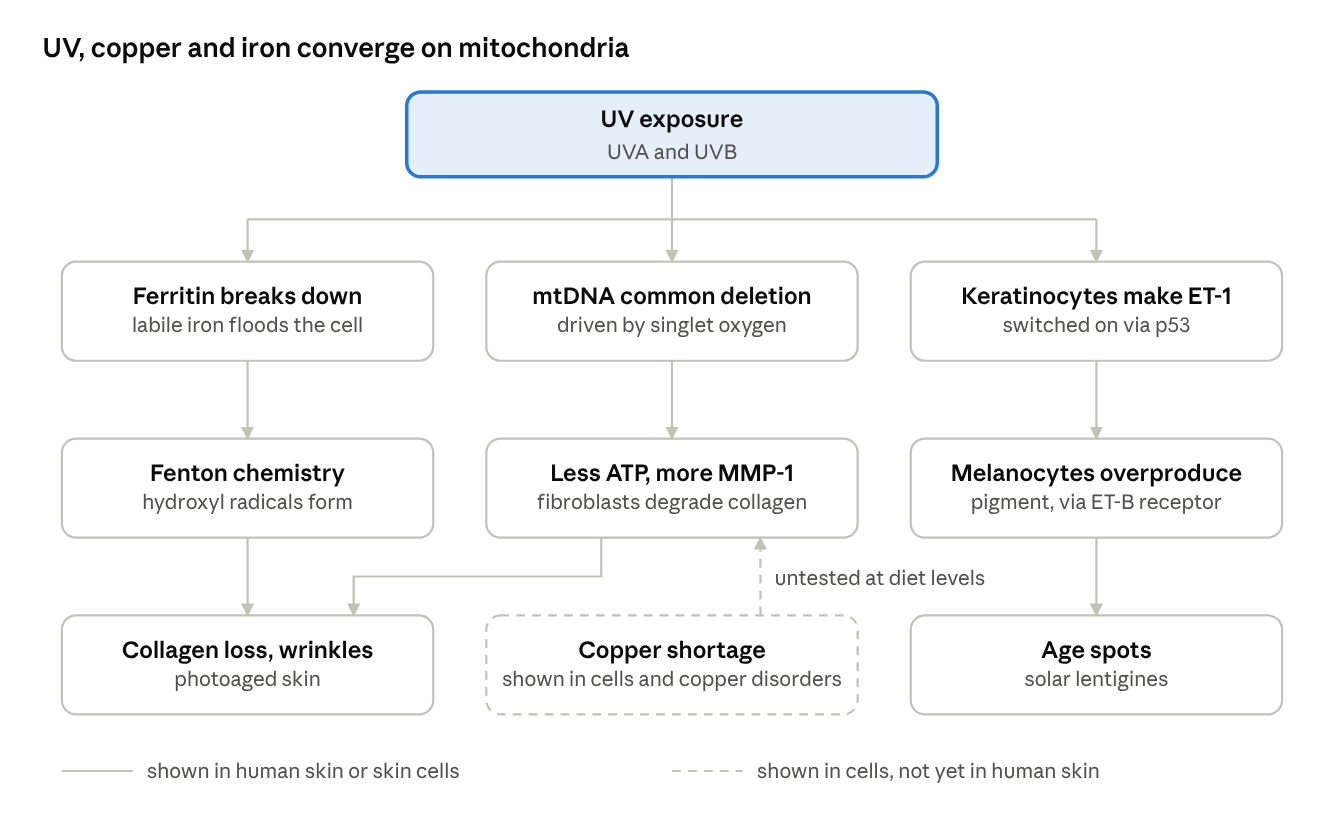
skin ageing pathways · graded by evidence
Each UV-driven step has been shown in human skin or human skin cells. The links popular protocols add (marginal copper intake starving mitochondria, iron accumulation switching on ET-1) have not been tested in people.
Blue light and the iron band
Blue light sits in the iron band. Iron-containing heme compounds, haemoglobin and the cytochromes, absorb most strongly in the blue region around 400 nm, the Soret band (optical properties of skin); haemoglobin peaks at 418 nm (Lim 2022). That is the same band that drives pigmentation:
- Sunlight builds an iron-type pigment in skin. Modest solar exposure produced an epidermal chromophore absorbing at about 412 nm, strong enough to tint skin yellow to reddish, whose peak strongly suggests a heme-like compound; the authors propose it contributes to the colour of chronically sun-exposed skin (Dowdy 2004).
- Blue light around 415 nm triggers lasting pigment. In melanocytes it acts through OPN3, producing long-lasting pigmentation in skin types III and above (Regazzetti 2018).
It loads mitochondria with oxidative stress. Blue light, but not green, red or infrared, caused mitochondrial oxidative stress in live skin, and the blue light in sunlight measurably affected human skin (Nakashima 2017).
Iron oxide blocks the band from outside. In melasma, iron-oxide sunscreen plus hydroquinone improved MASI scores more than a UV-only sunscreen plus hydroquinone (Castanedo-Cázares 2014).
In skin carrying extra iron, blue light falls on the very wavelengths iron compounds absorb while also switching on pigment through OPN3, which helps explain why visible light, not just UV, worsens melasma.
Pigmentation and melasma have their own post. How ET-1, iron, copper and light apply to melasma, sun spots and pigmentation after inflammation or procedures, with the practical steps, is in Pigmentation and Melasma: Copper, Iron, ET-1 and Light.
How the key claims stand in the research
Most of the claims central to the copper-iron view of skin have research behind them once you look past the headline studies. Each is graded below by the strongest evidence found, from human trials down to cell work.
| Claim | Evidence | What the research shows |
|---|---|---|
| High-dose synthetic ascorbic acid disturbs copper-iron handling | Supported: human and cell studies | 1,500 mg a day for 64 days lowered ceruloplasmin activity at every checkpoint, and serum copper rose significantly within 20 days of stopping, showing a direct, reversible effect of the supplement (Finley & Cerklewski 1983); 605 mg a day cut its oxidase activity by 21% (Jacob 1987). Dietary ascorbic acid reduced copper absorption in rats (Van den Berg 1992). In cells it expanded the loose iron pool (Bridges & Hoffman 1986) and, with iron, released ferritin iron at normal body concentrations (Badu- Boateng 2019). Overall human copper status was unchanged in the 605 mg study. |
| Ascorbic acid promotes fibrosis | Supported in cell culture | It amplified TGF-β1-driven myofibroblast changes and collagen deposition in human dermal fibroblasts (Piersma 2017). Topical vitamin C trials in photoaged skin have so far shown improvement (Humbert 2003). |
| High-dose synthetic vitamin D causes harm | Supported: randomised trials at high and bolus doses | 4,000–10,000 IU daily caused more bone loss and urine calcium than 400 IU (Calgary); a yearly 500,000 IU dose raised falls and fractures. Vitamin A blunted the calcium response to vitamin D in humans (Johansson & Melhus 2001). Frank calcification needs toxic levels (Vieth 2007), and 800 IU a day did not lower serum retinol (Chai 2012). |
| Whole-food vitamin C differs from ascorbic acid | Partly supported | Food brings flavonoids, polyphenols and lower doses, without the gram-dose effects above. The vitamin C molecule itself is identical and gave the same blood and tissue levels in human trials (Carr & Vissers 2013). I found no study showing a tyrosinase core within the vitamin C complex, and many acerola powders contain added ascorbic acid (example label). |
| Animal nutrients are more bioavailable | Largely supported | B12 is absent from unfortified plants, heme iron and pre-formed retinol are absorbed directly, and common BCMO1 variants cut beta-carotene conversion by 32-69% (Leung 2009). |
| Cacao carries heavy metals | Supported | Of 72 US cocoa products, 43% exceeded California's Prop 65 lead limit and 35% the cadmium limit (Hands 2024). The same team notes shellfish and organ meats can also accumulate heavy metals (GWU), so sourcing matters for those too. |
Heme iron and the balance. Heme iron from red meat is absorbed efficiently, which helps where iron is low but adds to stores where they are already high. Meat-rich eating pairs well with the iron-limiting levers in the priming section, such as tea with meals and checking ferritin.
Liver as food is not the same as vitamin A supplements. Liver is one of the richest sources of pre-formed retinol, but the body handles it differently from tablets: blood levels of vitamin A and its active metabolites were many-fold higher after a supplement than after a liver meal, and the author concluded that liver consumption does not increase the risk of birth defects (Honeywell 2001). The key study linking vitamin A to birth defects found the risk with supplements above about 10,000 IU a day (Rothman 1995), and there are no convincing cases of foetal harm from vitamin A in food (Bates 1995, via GPnotebook). The documented cases of liver-related vitamin A toxicity involve extreme, frequent intakes: infants fed 120 g of chicken liver every day for months, adults eating liver at several meals a day or more than 1 kg of meat and liver daily alongside supplements (Castaño 2006). No study has shown harm from eating liver as an ordinary food, such as a portion once a week; the problems documented with vitamin A come from supplementation and from extreme daily liver intakes. UK antenatal guidance still advises avoiding liver in pregnancy as a precaution.
Vitamin D and the skin
Vitamin D matters for skin, but the evidence favours correcting a real shortfall and avoiding excess, not routine high dosing. My companion post, The Case for Caution: Rethinking Synthetic Vitamin D, covers the systemic trials in depth; here is what bears on skin.
Sunlight's built-in brake, and the UK winter
Skin production of vitamin D self-limits: with longer exposure, surplus pre-vitamin D turns into inactive photoproducts, so sunlight alone does not cause toxicity. But the season limits it too. On cloudless days, skin in Boston (42°N) made no pre-vitamin D from November to February, and in Edmonton (52°N) from October to March (Webb 1988). Engelsen 2005, which covers the whole of the UK. Winter vitamin D has to come from summer stores, food and cod liver oil.
What vitamin D does in skin
The receptor matters for skin structure. Mice lacking the vitamin D receptor show premature ageing, including early hair loss, thickened skin, enlarged sebaceous glands and epidermal cysts (Keisala 2009). Much of this hair and keratinocyte role is ligand-independent: mice fed a vitamin D-free diet for five generations kept normal hair cycling (Bikle review). So a working receptor matters more for skin than pushing blood levels high.
Fibroblast effects (cell culture). Active vitamin D stimulated type I collagen, inhibited elastase activity and reduced oxidative damage in UVA-exposed human dermal fibroblasts (Philips 2019).
Sunburn inflammation (human). In a small randomised trial, 200,000 IU given an hour after experimental sunburn lowered skin TNF-α and nitric oxide synthase (Scott 2017, summarised). That was a single bolus, the dosing pattern linked to harm in older women, so it is a mechanism, not a recommendation.
Healing. Pooled trials in diabetic foot ulcers suggest supplementation in deficient patients improves healing, with an important caveat about bias in the included studies (umbrella review 2024).
Pigmentation. People with melasma had lower 25(OH)D in observational studies (Skin 2023), but no trial has shown supplementation improves melasma, and sun avoidance alone could explain the gap.
I found no randomised trial showing vitamin D supplements improve wrinkles or elasticity in people who are not deficient.
Where more becomes a problem
The cautions are real, and they come from randomised trials rather than theory:
- In the Calgary trial, 4,000 and 10,000 IU daily for three years produced more bone loss than 400 IU, alongside more high urine calcium.
- A single 500,000 IU yearly dose raised falls by 15% and fractures by 26% in older women.
- Vitamin A blunted the calcium response to vitamin D in a human study, so the two fat-soluble vitamins are best kept in balance. That said, 800 IU of vitamin D daily did not lower serum retinol over six months (Chai 2012), so a functional vitamin A deficiency from ordinary doses has not been shown.
- Vitamin D lowers hepcidin, which governs iron handling; the long-term effect on tissue iron, the subject of this post, has not been studied.
Practical take for skin. Get summer sun on arms and legs with the face protected, eat oily fish and eggs, and take cod liver oil through the winter rather than a vitamin D supplement. Interpret a 25(OH)D result alongside active vitamin D (1,25-dihydroxyvitamin D), PTH and calcium rather than chasing a high number. Active vitamin D does not measure stores: it can be normal or even raised when 25(OH)D is low, because a rising PTH drives the kidney to convert more. A high active vitamin D can also signal inflammation. Activated macrophages make their own 1,25-dihydroxyvitamin D, outside the kidney's PTH control; in granulomatous inflammation such as sarcoidosis or tuberculosis this can push active vitamin D and calcium up while PTH is suppressed (sarcoidosis review; Tebben 2016). So a high 1,25 with a low or normal 25(OH)D is a reason to look for inflammation, not to give more vitamin D. High active vitamin D also uses up stored vitamin D. In people given tracer-labelled 25(OH)D, raising 1,25-dihydroxyvitamin D significantly shortened the half-life of 25(OH)D, within 24 hours of starting, and the authors concluded this accelerated breakdown contributes to vitamin D deficiency in many clinical disorders (Clements 1992). Inflammation lowers the stored form directly too: after knee replacement, 25(OH)D fell by 40% within two days as CRP rose and was still 20% lower three months later, leading the authors to conclude that 25(OH)D is not a reliable measure of vitamin D status when there is significant inflammation (Reid 2011). So a low stored vitamin D can be a result of inflammation, with high active vitamin D speeding its breakdown, rather than simply a lack of sun or food; check CRP before reading a low 25(OH)D as deficiency. Read together, the four show how the body is actually handling vitamin D.
Ascorbic acid in depth
Ascorbic acid is one of the best-studied nutrients in skin biology, and the evidence points one way: keep skin saturated, use topical forms correctly, and skip megadoses. The fears about copper loss and fibrosis come from real lab chemistry, applied to situations where it does not hold.
How much reaches the skin
Vitamin C is pumped into cells by two transporters, SVCT1 and SVCT2, so uptake is tightly regulated. Classic depletion-repletion work by Levine and colleagues (PNAS 1996) showed plasma levels plateau at intakes of around 200 mg a day; at gram doses, the fraction absorbed falls and the excess is excreted in urine.
Skin tracks blood closely. In a 2025 study from Otago, dermal fibroblasts held roughly seven times more vitamin C than epidermal cells, and skin levels rose in proportion to plasma. Twenty-four adults who ate two gold kiwifruit a day (about 250 mg vitamin C) for eight weeks reached plasma saturation. Their skin density rose from about 0.15 to 0.23 scanner units, and the outer skin renewed faster (Pullar 2025). The honest caveats: it was small and uncontrolled, published as a letter, elasticity dipped slightly, and a procollagen marker in blister fluid did not rise (summary).
At population level, among 4,025 American women, higher dietary vitamin C was linked to lower odds of a wrinkled appearance and of age-related dryness, after adjusting for sun exposure, smoking and other factors (Cosgrove 2007). That study was cross-sectional and led by Unilever scientists, so it shows association, not cause.
What it does in skin
- Collagen hydroxylation. Prolyl and lysyl hydroxylases need vitamin C to keep their iron atom in the active ferrous state. Without it, collagen chains cannot form stable triple helices, which is why scurvy wounds reopen. This is vitamin C putting iron to productive use, not a hazard.
- Epigenetic enzymes. The TET DNA demethylases and several histone demethylases also depend on vitamin C, which is how it switches on whole gene programmes in connective tissue cells (Thaler 2022).
- Antioxidant recycling. It scavenges radicals in water-based compartments and regenerates vitamin E in membranes.
- Pigment. Ascorbic acid inhibits melanin production; ascorbyl phosphate is used as a skin-lightening agent in age-spot research (Imokawa 2019). This is the opposite of the claim that "real" vitamin C carries a tyrosinase core.
Is it a pro-oxidant in the body?
In a test tube, ascorbate plus free iron or copper generates hydroxyl radicals. The question is whether that happens in people, where iron is almost entirely bound to transferrin and ferritin. Carr and Frei reviewed the evidence and found compelling antioxidant protection of lipids by vitamin C in biological fluids, animals and humans, including when iron was given at the same time; DNA findings were inconsistent and often methodologically flawed (Carr & Frei 1999). Even in plasma from iron-overloaded patients, vitamin C behaved as an antioxidant (Berger 1997, J Biol Chem).
The real exception is absorption, not oxidation: vitamin C increases uptake of non-heme iron from the gut. People with haemochromatosis or other iron overload should avoid vitamin C supplements taken with meals.
Where the pro-oxidant concern is real. Ascorbate does become a pro-oxidant when genuinely free iron is present, and some people have it. In severe iron overload, such as thalassaemia or advanced haemochromatosis, transferrin is saturated and non-transferrin-bound iron circulates. That is why vitamin C given with the chelator deferoxamine in transfusion-overloaded patients was linked to worsening heart function (Nienhuis 1981, NEJM), and why it is now introduced cautiously, at low dose, in that setting. The same logic applies inside a skin cell in the minutes after UVA releases labile iron (free iron) from ferritin. But that is a reason to block the UVA, not evidence that dietary vitamin C ages skin: in pig skin, topical vitamin C combined with vitamin E reduced UV damage (Lin 2003, J Am Acad Dermatol).
Inside the cell: ascorbate, ferritin and the labile iron (free iron) pool
The plasma studies above measure blood. Inside cells, where iron is stored, the picture is less reassuring, and it bears directly on iron accumulation.
Ascorbate holds iron in ferritin but swells the free pool. In human cells, ascorbate slowed the breakdown of ferritin in lysosomes and stabilised its stored iron, yet the pool of chelatable (loosely held) iron expanded (Bridges & Hoffman 1986). The same group summarised the consequence: more "free" iron within cells, capable of damaging lipids, proteins and DNA (NIH grant summary).
Ascorbate and iron together release iron from ferritin. A 2019 review showed that small amounts of loose iron accelerate ascorbate's ability to dissolve ferritin's iron core, lowering the ascorbate concentration needed into the normal physiological range (50-250 µM). The authors note that a rise in the labile iron (free iron) pool could then tip cells into ferroptosis, with relevance to inflammatory conditions (Badu-Boateng & Naftalin 2019).
In ferroptosis research, the pro-oxidant side tends to win. A 2024 review in Signal Transduction and Targeted Therapy notes that in the few ferroptosis studies so far, vitamin C's pro-oxidant role predominated over its antioxidant one (In defence of ferroptosis). Most of this work used cancer cells and high or intravenous doses, which raise the labile iron (free iron) pool deliberately.
- Ascorbate also moves iron out. In human gut cells, intracellular ascorbate increased ferroportin, the only known iron exporter (Scheers 2014), so its net effect depends on cell type and iron status.
What this means for skin with iron accumulation. The cell biology gives a coherent reason for caution with high-dose ascorbic acid where iron is already building up: ascorbate can enlarge the loose iron pool that feeds the Fenton reaction, and this happens at concentrations reached in the body. It has not been tested in human skin, and topical vitamin C in human studies has so far been protective (see below). The practical line is food-level vitamin C, no high-dose supplements when ferritin is raised, and not taking vitamin C supplements with iron-rich meals in that situation.
Ascorbate sits at the copper-iron crossroads
The gut enzyme that prepares dietary iron for absorption, duodenal cytochrome b (Dcytb), uses ascorbate inside the cell to reduce ferric iron to ferrous iron (Ganasen 2018). The same enzyme also reduces copper: cells over-expressing Dcytb took up 6.7 times more copper, leading researchers to describe it as a dual iron and copper reductase (Solomou 2013). Ascorbate also multiplied copper transfer from ceruloplasmin into cells at least fourfold, while not increasing copper loading into the antioxidant enzyme SOD (Percival & Harris 1989). So vitamin C is not an isolated nutrient either: it is one of the reducing agents the copper-iron axis runs on.
How large is the iron-absorption effect? Added to a single meal, ascorbic acid reliably increases non-heme iron absorption, and a meta-analysis found long-term supplementation raised haemoglobin (Heffernan 2017). Across a whole mixed diet, though, vitamin C intakes from 51 to 247 mg a day made no significant difference to absorption (Cook & Reddy 2001). The effect is real, largest with supplements taken alongside iron, and smaller with ordinary eating.
The copper question, both directions
High-dose ascorbic acid modestly lowers ceruloplasmin oxidase activity in humans without changing copper absorption or status (see the myth table). In rats, dietary ascorbic acid reduced intestinal copper absorption, yet it also increased the efficiency of copper uptake into liver cells (Van den Berg 1992). The cellular copper importer CTR1 only carries reduced copper (Cu+), and ascorbate is one of the reductants that makes this possible; copper uptake and total cellular copper rise in its presence (Schwab 2016). "Strips copper from tissues" is not an accurate summary.
Ascorbate and ceruloplasmin, precisely. The effect on ceruloplasmin is real but specific. In the test tube, ascorbate inhibits ceruloplasmin's ferroxidase activity (Løvstad 1997), and plasma ascorbate blunts its antioxidant activity (Gutteridge 1991, Clin Sci), partly because ceruloplasmin oxidises ascorbate itself and simply re-reduces the iron it has just oxidised. In people, 605 mg a day cut measured oxidase activity by 21% while ceruloplasmin protein, copper absorption and copper status stayed unchanged (Jacob 1987), and vitamin C at 100 mg a day or more interferes with the oxidase assay itself, so some of the measured drop is laboratory artefact (Metabolism 1995). What has never been shown is the next step the popular argument needs: that this measured change leads to iron building up in human tissues, including skin.
Does it cause fibrosis?
This claim has more behind it than I first credited. In a peer-reviewed Groningen study, ascorbic acid did more than support collagen production in human dermal fibroblasts. Working with the pro-fibrotic signal TGF-β1, it amplified collagen I and IV deposition, α-smooth-muscle-actin stress fibres, contraction of collagen gels and several fibrosis-linked genes: a genuine push towards the myofibroblast phenotype (Piersma 2017). The undergraduate thesis often cited for this claim (Seijkens 2015) came from the same research group.
Three limits matter. First, the effect needed TGF-β1, an injury signal, to be present: ascorbate amplified an existing fibrotic drive rather than starting one. Second, human fibroblasts cultured without vitamin C are effectively scorbutic, so "adding" it in a dish models going from deficient to normal, not from normal to high. Third, no human study shows dietary or topical vitamin C causing skin fibrosis, while randomised trials of topical vitamin C show improved, more elastic photoaged skin. A fair summary: in skin that is already wounded or inflamed, vitamin C helps whatever repair programme is running, whether that is healthy remodelling or scarring.
Does pure ascorbic acid cure scurvy?
A popular claim says Albert Szent-Györgyi, who isolated the molecule, believed ascorbic acid does not cure scurvy. The record shows something narrower. In 1932 he and Joseph Svirbely showed that 1 mg a day of the purified molecule protected guinea pigs from scurvy and declared it identical to vitamin C (NLM archive). In 1936 he went further and proposed that citrus flavones ("vitamin P") were needed for capillary strength, arguing guinea-pig scurvy was a combined C and P deficiency; Zilva could not confirm this, and Szent-Györgyi himself did not reproduce it (1941 summary). His later recollection that pure vitamin C helped a bleeding patient less than his crude extract concerned capillary bleeding, not scurvy as a whole.
Human experiments settled the question. In the Iowa studies, men fed a vitamin C-free diet developed scurvy within about three months; purified ascorbic acid reversed it, with as little as 6.5 mg a day curing the clinical signs in one man and roughly 10 mg a day sufficient to prevent or cure it (Hodges 1969; Hodges 1971). The fair kernel: dietary flavonoids may add their own small benefit to capillary resistance, which is a reason to eat fruit, not a reason to fear ascorbic acid.
Getting topical vitamin C to work
In pig skin, L-ascorbic acid entered only when formulated below pH 3.5, absorption peaked at 20% and fell at higher strengths, skin levels saturated after three daily applications, and they halved over about four days after stopping. In that study, magnesium ascorbyl phosphate and ascorbyl palmitate did not raise skin L-ascorbic acid (Pinnell 2001). Note that Pinnell's formulation became a commercial serum. L-ascorbic acid oxidises quickly, turning yellow-brown; opaque, airless packaging matters, and the low pH can sting sensitive skin. On human skin exposed to solar-simulated UV, a 15% L-ascorbic acid, 1% vitamin E and 0.5% ferulic acid serum reduced redness, sunburn cells, thymine-dimer DNA damage and inflammatory cytokines versus its vehicle (Murray 2008); the study was small and funded by the manufacturer, and adding ferulic acid had doubled photoprotection in earlier pig-skin work (Lin 2005).
Safety
- Kidney stones. Among 23,355 Swedish men followed for 11 years, those taking vitamin C tablets (usually 1,000 mg) had about twice the rate of kidney stones; multivitamins showed no link (Thomas 2013). Part of absorbed vitamin C is excreted as oxalate.
- Iron overload: avoid supplements, as above.
- High-dose intravenous vitamin C can cause haemolysis in G6PD deficiency and oxalate kidney injury in renal impairment; screen before "drips".
Practical take
- Aim for plasma saturation, roughly 200-250 mg a day, which food can supply: two kiwifruit, a red pepper, or a portion of broccoli plus citrus.
- Food and tablets deliver the same molecule; gram doses add stone risk, not skin benefit.
- For a topical, look for L-ascorbic acid at 10-20% and low pH in light-proof packaging, used alongside sunscreen.
For skin, high doses add nothing. Skin takes its vitamin C from the blood, and skin levels track plasma levels more tightly than in any other organ the Otago group has studied (Pullar 2025). Plasma, in turn, has a ceiling. In carefully controlled depletion–repletion studies, 200 mg a day brought plasma to more than 80% of its maximum with tissues saturated; at single doses of 500 mg and above, absorption fell and the extra was excreted, and the authors concluded that daily doses above 400 mg have no evident value (Levine 1996; NIH release). The skin benefits seen in the kiwifruit study came from about 250 mg a day from food, which raised skin vitamin C, skin density and epidermal renewal in people starting from low levels (Pullar 2025). So once plasma is saturated, a high dose cannot push more vitamin C into skin; it only adds the costs above, lower ceruloplasmin activity, more loose iron and more oxalate.
The practical takeaway is to avoid chronic high-dose vitamin C supplements, particularly where copper status is low or iron is accumulating. Food sources don't reach the doses that lowered ceruloplasmin activity in human trials.
Food comes out ahead of supplements because of what travels with the vitamin C, fibre, polyphenols and the rest of the food matrix, not because the vitamin C molecule itself is different.
If you do choose a supplement, check purity. Choose a reputable brand whose ascorbic acid meets pharmacopoeial grade (BP, Ph. Eur. or USP). The European Pharmacopoeia monograph caps copper at 5 ppm and iron at 2 ppm, which matters for the copper-iron balance, and also limits oxalic acid to 0.2%; suppliers' pharmacopoeial-grade specifications add limits for lead, arsenic, mercury and other heavy metals (VWR specification). Keep the dose modest, for the reasons above.
Four more drivers that fit the same picture
Nicotinamide: energy for DNA repair
Nicotinamide, the amide form of vitamin B3, is a precursor of NAD+, the coenzyme mitochondria need to make ATP. UV drains keratinocytes of energy, and nicotinamide prevented that UV-induced ATP decline in human keratinocytes; it also increased DNA repair and reduced both UV-induced dimers and oxidative DNA damage in keratinocytes and in human skin samples (Surjana 2013). That is the mitochondria section in practice: DNA repair is energy-hungry, and a cell short of ATP repairs less.
The human trial is strong. In ONTRAC, 386 people who had had at least two non-melanoma skin cancers in five years took 500 mg of nicotinamide twice daily or placebo for 12 months; new non-melanoma skin cancers fell by 23% (Chen 2015, NEJM). The benefit was no longer seen once people stopped taking it (MDedge report). Two cautions: the trial was in a high-risk group, and the investigators did not recommend it for the general population (MDedge); and nicotinamide is not niacin (nicotinic acid), which causes flushing. For everyone else, the food route is protein: meat, fish and poultry supply both niacin and tryptophan, from which the body makes NAD+.
Glycation: sugar stiffening collagen
Collagen is long-lived, so it collects chemical damage. Sugars attach to it (glycation) and, with oxidation, form cross-linking "glycoxidation" products such as carboxymethyl-lysine (CML) and pentosidine. In human skin collagen, the initial glycation product rose only 33% between ages 20 and 85, but CML, pentosidine and collagen fluorescence rose about fivefold with age; in diabetes, glycation was three times higher and tracked HbA1c (Dyer 1993, JCI). The authors described diabetes as accelerated chemical ageing of long-lived proteins. Because the damaging products need oxidation as well as sugar, the same oxidative environment this post describes, free iron, weak Complex IV, low SOD1, would be expected to add to this damage, although that link has not been measured directly in skin. Practically: keep blood glucose steady and know your HbA1c.
Omega-3 fatty acids: a raised sunburn threshold
In a double-blind trial, 42 people took 4 g a day of the omega-3 EPA or oleic acid for three months. On EPA, the UV dose needed to cause redness rose from 36 to 49 mJ/cm2, and UV-induced p53 in skin, a marker of DNA damage, halved; oleic acid did nothing (Rhodes 2003). An earlier fish-oil study more than doubled the burning threshold over six months, though skin lipid peroxidation rose, suggesting omega-3s act as a sacrificial buffer (Rhodes 1994). Recommended: oily fish and cod liver oil. Oily fish is the food route, and cod liver oil takes the place of omega-3 or fish-oil capsules. Cod liver oil is the traditional whole-food concentrate and brings three things this post keeps returning to in one spoon: the omega-3s EPA and DHA, pre-formed vitamin A for the skin's light sensing and iron handling, and vitamin D. The 2003 trial used isolated, purified EPA; this post suggests cod liver oil instead, the whole-food form, which supplies EPA and DHA together as the fish-oil study did, alongside vitamins A and D. Products vary in their vitamin A and D content, so check the label; with cod liver oil as the winter source, no separate vitamin D supplement is needed.
Air pollution: pigment spots beyond the sun
In 400 German women aged 70-80, higher exposure to traffic soot and particles was associated with 20% more pigment spots on the forehead and cheeks, and less strongly with wrinkles (Vierkötter 2010); the finding was later replicated in Caucasian and Asian cohorts (review). Polycyclic aromatic hydrocarbons on the particles act through the aryl hydrocarbon receptor, which also stimulates melanocytes, and carbon nanoparticles can penetrate skin (MDedge; Cosmetics Design). The proposed mechanism is oxidative stress, the same pathway as the rest of this post.
Priming from the inside: food first, light, targeted support
Priming means rebalancing the copper-iron axis and protecting mitochondria before asking fibroblasts to rebuild. Each step below acts on that axis and is backed by primary research.
1. Food first: feed copper, manage iron
Copper-rich whole foods. Beef liver, oysters and other shellfish, nuts and seeds supply copper in food form, avoiding the gut competition caused by high-dose zinc or iron supplements. In rodents, iron loading caused copper deficiency and extra dietary copper prevented it (Ha 2016).
Time polyphenol drinks with meals to limit non-heme iron uptake. In human absorption studies, black tea with a meal cut non-heme iron absorption by 79–94%, peppermint tea by 84%, cocoa by 71% and chamomile by 47% (Hurrell 1999). Coffee works the same way through its chlorogenic acids: a cup with a hamburger meal cut iron absorption by 39% (tea cut it by 64%), stronger coffee cut it further, and the effect was concentration-dependent. Timing matters: coffee drunk an hour before a meal had no effect, while coffee drunk an hour after inhibited absorption as much as drinking it with the meal (Morck 1983). For someone with high ferritin, tea or coffee with or after meals is a simple lever; for someone iron-deficient, it is the opposite, and the drink is better taken at least an hour before eating.
Vitamin C from food, not gram-dose tablets, for the reasons in the ascorbic acid section, especially where ferritin is raised.
Protein and glycine-rich foods for collagen building blocks. Meat, fish, eggs and dairy supply complete protein; glycine, which makes up about a third of the amino acids in collagen, is richest in collagen-containing foods such as bone broth, slow-cooked cuts with connective tissue, poultry skin, fish skin and gelatin. Animal foods also bring heme iron, zinc, copper, B12 and pre-formed vitamin A in well-absorbed forms. Oral collagen peptides as supplements are uncertain: a 2023 meta-analysis found benefit (Nutrients 2023), but a 2025 analysis found none in high-quality or independently funded trials (summary).
Magnesium-rich foods daily. Pumpkin and other seeds, nuts, dark leafy greens, legumes and cacao supply the magnesium mitochondria need to make usable ATP. Intakes are commonly low: more than half of the US population falls below the Estimated Average Requirement, and moderate magnesium deficiency accelerated ageing in human fibroblasts (Killilea & Ames 2008). Phytates in grains and legumes bind magnesium, so soaking or sprouting them helps.
Oily fish and cod liver oil for omega-3s. EPA raised the skin's sunburn threshold and halved UV-induced p53 in a randomised trial (Rhodes 2003). Cod liver oil adds pre-formed vitamin A and vitamin D to the omega-3s; it is this post's winter source of vitamin D, in place of vitamin D supplements. Check the label, as content varies by product.
2. Lower stored iron where it is high
Blood donation. Frequent donors had median ferritin of 17 versus 52 ng/mL in infrequent donors, lower 3-nitrotyrosine (an oxidative-stress marker) and better artery function (Zheng 2005). In 150 donors, more donations meant lower ferritin, lower malondialdehyde and higher SOD activity (Mehrabani 2008). These are observational, and donation is only appropriate where ferritin is genuinely high.
Curcumin as a food-derived iron chelator. In mice, dietary curcumin reduced liver and spleen iron and suppressed hepcidin and ferritin (Jiao 2009); over six months it lowered iron stores without lowering zinc or copper (Huebbe 2014). The flip side: it can tip low-iron people into deficiency, so check ferritin first.
Polyphenols that protect stored iron. Epicatechin prevented UVA from releasing iron in skin fibroblasts by protecting lysosomes (Basu-Modak 2006).
3. Light: regular, well-timed sun, not avoidance
Avoiding the sun does not create healthy skin. Skin is built to use sunlight, and low, regular doses make it stronger:
It strengthens the barrier. Suberythermal UVB (about half a burning dose, daily for three days) accelerated barrier recovery and raised the differentiation proteins involucrin and filaggrin along with the skin's antimicrobial peptides; blocking the skin's vitamin D activation abolished these benefits (Hong 2008). In 258 people, barrier recovery was delayed only at higher doses of sun exposure (2010 study).
It builds the skin's own protection. A simulated summer of low-level sunlight in light-skinned people increased melanin and thickened the stratum corneum from 29 to 41.5 μm, giving measurable, if modest, photoprotection even in very light skin (Felton 2020). In another study, repeated low doses raised melanin by 12%, stratum corneum thickness by 34% and vitamin D by 21% (RIVM 2025).
It supplies what skin cannot get the same way elsewhere: vitamin D from UVB, nitric oxide from UVA, and the red and near-infrared light that Complex IV absorbs.
The whole-body picture points the same way. In 29,518 Swedish women followed for 20 years, those who avoided the sun had about twice the mortality of those with the most sun exposure (Lindqvist 2014). The authors concluded that very restrictive sun advice in low-sunlight countries may harm women's health; the study is observational, so lifestyle differences may explain part of it.
The line is dose, not avoidance. Even suberythermal UV leaves some DNA damage that adaptation then limits, and burning doses drive the mitochondrial DNA and collagen damage described earlier. So the goal is regular, moderate, well-timed exposure that never reaches burning, with the face shaded at peak UV.
Red and near-infrared light act on the copper centres of Complex IV. Karu's action spectra showed that cytochrome c oxidase, with its two copper and two heme centres, is the main absorber of red and near-infrared light in cells (Hamblin review). In a randomised controlled trial of 136 volunteers, 30 sessions of red or red/near-infrared light improved skin roughness, wrinkles and ultrasound-measured collagen density versus controls (Wunsch & Matuschka 2014).
Limit the iron and ET-1 triggers. UVA releases labile iron (free iron) from ferritin and UVB switches on keratinocyte ET-1 (sections above). In the Nambour trial, 903 adults randomised to daily broad-spectrum sunscreen showed 24% less skin ageing over 4.5 years (Hughes 2013). Infrared-A in strong sunlight also raises MMP-1 in human skin (Krutmann), so the face benefits from shade even when arms and legs get sun for vitamin D.
4. Movement for blood flow and ET-1
Three months of aerobic exercise reduced ET-1-driven vasoconstriction in previously sedentary older men (Van Guilder 2007), improving the blood supply that dermal fibroblasts depend on.
5. Targeted topicals
Copper peptides (GHK-Cu) and copper-oxide textiles deliver copper to the skin surface, with small positive trials (Borkow 2009).
Topical iron chelators reduced UV wrinkling in animals and UV redness in a human study (Bissett 1994); kojic acid is one cosmetic ingredient with chelating activity (Mitani 2001).
Topical L-ascorbic acid with vitamin E and ferulic acid reduced UV damage in human skin (Murray 2008).
6. Test before you treat
A panel that maps the axis: ferritin and transferrin saturation, serum copper and ceruloplasmin (oxidase activity where available), zinc, red blood cell (RBC) magnesium, vitamin D (25(OH)D and active 1,25-dihydroxyvitamin D) with PTH and calcium, full blood count, and CRP to interpret the rest. The same panel is set out in the vitamin D post.
Why RBC magnesium rather than serum magnesium. Less than 1% of the body's magnesium is in the blood, and serum levels are held steady by drawing on bone stores, so a normal serum result does not rule out moderate to severe deficiency. RBC magnesium measures magnesium inside cells and often reflects body status better than serum (Razzaque 2018; DiNicolantonio 2018). It is still not a perfect marker, since no single blood test captures tissue magnesium fully, so read it alongside diet and symptoms.
Not supported by the research so far: gram-dose vitamin C where ferritin is raised, high-dose or bolus vitamin D for skin, long-term high-dose zinc without copper, and iron supplements without a confirmed deficiency.
Inflammatory skin conditions
The same copper-iron, ET-1 and nitric oxide systems run through eczema, psoriasis and dermatitis, where inflammation also shifts the blood markers. That has its own post: Eczema, Psoriasis and Dermatitis: The Copper-Iron Axis in Inflamed Skin.
Does inside-out priming improve treatment results?
The logic is sound, since every treatment depends on the patient's own fibroblasts, but no trial has tested a nutritional "priming" protocol before an aesthetic procedure. What we have is indirect evidence.
What we do know. Malnutrition and specific deficiencies worsen healing, scarring and pigmentation after aesthetic and reconstructive procedures (Vitagliano 2023; Seth 2024). Smoking and alcohol impair wound repair. So correcting a genuine deficiency, and avoiding procedures during a crash diet or illness, is well grounded. Whether going from adequate to optimised nutrition improves outcomes has not been studied.
Collagen stimulators (PLLA, CaHA). PLLA works through a controlled foreign-body response: macrophages shift to a repair (M2) phenotype and release TGF-β, which drives fibroblast collagen synthesis (Avelar 2024). In a small randomised, placebo-controlled study of 10 women, PLLA produced histological rejuvenation 18 weeks after the last session (Arruda 2024). Encouragingly for older patients, PLLA still increased collagen and elastin in aged mouse skin and reduced MMP-2 and MMP-3 (Oh 2023). There is no evidence that marginal copper status blunts this response in practice; the theoretical concern is only relevant to true deficiency.
PRP. The problem with PRP is not "starved plasma"; it is that the overall evidence is weak. An overview of 13 systematic reviews rated confidence low or critically low in 12, and concluded the evidence is insufficient for firm conclusions about facial rejuvenation (Cruciani 2024). Preparation methods vary so much that platelet counts were reported in only a handful of studies (Xiao 2021). Until preparation is standardised, any effect of nutritional status is untestable.
HIFU and microfocused ultrasound. CFU-elite is one of these focused-ultrasound platforms. They heat tiny zones in the deep dermis and the SMAS above 60 °C, causing immediate collagen contraction and later remodelling. A 2025 systematic review reported laxity improvements of 18-30%, mostly in the lower face and neck, with transient side effects in under 5% (ASJ 2025). The evidence level is modest (mostly uncontrolled studies).
Post-inflammatory hyperpigmentation. The idea that iron-loaded skin and high ET-1 make lasers and microneedling more likely to cause PIH is a reasonable hypothesis that has not been tested. What has been tested surprises many clinicians: in a randomised trial of 100 CO2-laser patients, pretreatment with hydroquinone plus tretinoin, or glycolic acid, did not reduce post-laser hyperpigmentation compared with no pretreatment (West & Alster 1999). Strict sun avoidance before and after, appropriate energy settings for skin type, and early treatment if PIH appears have better support.
Bottom line for practice. Screen for and correct deficiencies, make sure protein intake is adequate, stop smoking, and protect from UV for weeks either side of a procedure. Be honest with clients that dietary "priming" beyond that is plausible but unproven.
At a glance: what works, what might, what doesn't
| Strategy | Evidence in humans | Verdict |
|---|---|---|
| Daily broad-spectrum sun protection | Large 4.5-year randomised trial | Works |
| Correcting true deficiencies (iron, B12, vitamin D, zinc, copper) before procedures | Strong for healing in general; untested for aesthetic outcomes specifically | Works where a deficiency exists |
| Adequate protein and vitamin C | Wound-healing literature | Works |
| Topical L-ascorbic acid | Randomised trials, modest effects | Works, modestly |
| Oral collagen peptides | Conflicting meta-analyses; funding bias | Uncertain |
| Topical copper peptides, copper textiles | Small trials with conflicts of interest | Promising |
| Topical iron chelators | Strong in mice; limited human data | Promising |
| Dietary polyphenols for skin iron | Cell studies only | Unproven |
| PRP for facial rejuvenation | Low-quality evidence overall | Unproven |
| Liver/oyster "priming" protocols | No trials | Unproven |
| Vitamin C from food rather than high-dose ascorbic acid | Same blood levels; high doses alter copper and iron handling | Works |
| High-dose (4,000 IU+) or bolus vitamin D | Randomised trials show more bone loss, falls and fractures | Avoid |
| Red and near-infrared light (photobiomodulation) | Randomised trial of 136 people; acts on Complex IV copper centres | Works |
| Tea, coffee or other polyphenol drinks with meals to limit iron uptake | Human absorption studies: 39– 94% less non-heme iron absorbed | Works where ferritin is high |
| Blood donation to lower high iron stores | Observational: lower ferritin and oxidative-stress markers in frequent donors | Promising |
| Regular aerobic exercise to lower ET-1 tone | Human intervention study in older men | Works |
| Regular, moderate, well-timed sunlight without burning | Barrier, photoadaptation and nitric oxide studies; large observational cohort | Works |
| Magnesium-rich foods; magnesium bath salts for the barrier | Controlled trials of magnesium-rich salt in atopic and psoriatic skin; fibroblast studies | Promising |
| Oral nicotinamide after previous skin cancers | Phase 3 randomised trial: 23% fewer new non-melanoma skin cancers | Works in high-risk groups |
| Oily fish and cod liver oil for omega-3s and the sunburn threshold | Randomised trial: higher burning threshold, less UV-induced p53 | Promising |
| Correcting low vitamin D in eczema (sunlight, cod liver oil) | Meta-analyses of randomised trials, mostly positive | Promising |
| Selenium supplements | Randomised trial: about 50% more type 2 diabetes | Avoid |
Safety cautions:
- High ferritin: check for hereditary haemochromatosis; avoid iron and high-dose vitamin C supplements, which increase iron absorption.
- High-dose vitamin C (ascorbic acid) supplements: add nothing for skin above about 400 mg a day and can disturb copper and iron handling (see the ascorbic acid section). Food-level vitamin C is not the concern.
- Zinc supplements: long-term high doses are a leading reversible cause of copper deficiency.
- Green tea extract: concentrated EGCG supplements at 800 mg a day or more carry a liver-injury signal.
- Shellfish and dark chocolate: both can carry heavy metals; variety and moderation beat any single "superfood".
Key sources
Iron and UV
- Killilea DW et al. Iron accumulation during cellular senescence in human fibroblasts in vitro. Antioxid Redox Signal 2003. Link
- Pourzand C et al. UVA induces immediate release of iron in human primary skin fibroblasts: the role of ferritin. PNAS 1999. Link
- Bissett DL, McBride JF. Synergistic topical photoprotection by an iron chelator and sunscreen. J Am Acad Dermatol 1996. Link
- Pelle E et al. Menopause increases the iron storage protein ferritin in skin. J Cosmet Sci 2013. Link
Copper
- Altarelli M et al. Copper deficiency: causes, manifestations, and treatment. Nutr Clin Pract 2019. Link
- Finley EB, Cerklewski FL. Ascorbic acid supplementation and copper status in young men. Am J Clin Nutr 1983. Link
- Jacob RA et al. Varying ascorbic acid intakes, copper absorption and ceruloplasmin. J Nutr 1987. Link
Mitochondria and ET-1
- Berneburg M et al. Induction of the photoaging-associated mitochondrial common deletion in vivo in human skin. J Invest Dermatol 2004. Link
- Berneburg M et al. Creatine supplementation normalizes mutagenesis of mitochondrial DNA. J Invest Dermatol 2005. DOI
- Imokawa G. Melanocyte activation mechanisms and treatments of solar lentigos. Int J Mol Sci 2019. Link
Vitamins C, A and D
- Carr AC, Vissers MCM. Synthetic or food-derived vitamin C: are they equally bioavailable? Nutrients 2013. Link
- Humbert PG et al. Topical ascorbic acid on photoaged skin: double-blind study vs placebo. Exp Dermatol 2003. Link
- Leung WC et al. BCMO1 polymorphisms alter beta-carotene metabolism. FASEB J 2009. Link
- Sha S et al. Safety of vitamin D supplements in 445,493 UK Biobank participants. Nutrients 2024. Link
Prevention and treatments
- Hughes MCB et al. Sunscreen and prevention of skin aging: a randomized trial. Ann Intern Med 2013. Link
- Hands JM et al. Heavy metal analysis of 72 dark chocolate and cocoa products. Front Nutr 2024. Link
- Cruciani M et al. PRP for facial rejuvenation: an overview of systematic reviews. Blood Transfus 2024. Link
- Seth I et al. Impact of nutrition on skin wound healing and aesthetic outcomes. JPRAS Open 2024. Link
- West TB, Alster TS. Pretreatment and hyperpigmentation after CO2 laser resurfacing. Dermatol Surg 1999. Link
Mitochondrial nutrients, nitric oxide and sunlight
- Acin-Perez R et al. Control of oxidative phosphorylation by vitamin A illuminates a fundamental role in mitochondrial energy homoeostasis. FASEB J 2010. Link
- Yamanaka R et al. Mitochondrial Mg2+ homeostasis decides cellular energy metabolism and vulnerability to stress. Sci Rep 2016. Link
- Killilea DW, Ames BN. Magnesium deficiency accelerates cellular senescence in cultured human fibroblasts. PNAS 2008. Link
- Liu D et al. UVA irradiation of human skin vasodilates arterial vasculature and lowers blood pressure independently of nitric oxide synthase. J Invest Dermatol 2014. Link
- Kourembanas S et al. Nitric oxide regulates the expression of vasoconstrictors and growth factors by vascular endothelium. J Clin Invest 1993. Link
- Hong SP et al. Biopositive effects of low-dose UVB on epidermis. J Invest Dermatol 2008. Link
- Felton SJ et al. Photoprotection conferred by low level summer sunlight exposures. Photochem Photobiol Sci 2020. Link
- Lindqvist PG et al. Avoidance of sun exposure is a risk factor for all-cause mortality. J Intern Med 2014. Link
- Rhodes LE et al. Recommended summer sunlight exposure levels can produce sufficient vitamin D at UK latitudes. J Invest Dermatol 2010. Link
- Levine M et al. Vitamin C pharmacokinetics in healthy volunteers. PNAS 1996. Link
- Pullar JM et al. Improved human skin vitamin C levels and skin function after dietary intake of kiwifruit. J Invest Dermatol 2025. Link
Liver and vitamin A
- Honeywell RJ. The pharmacokinetics of vitamin A in relation to its teratogenicity in healthy women. PhD thesis, University of Southampton, 2001. Link
- Rothman KJ et al. Teratogenicity of high vitamin A intake. N Engl J Med 1995. Link
- Castaño G et al. Vitamin A toxicity in a physical culturist patient: case report and review. Ann Hepatol 2006. Link
Inflammatory skin conditions
- Vähävihu K et al. Narrowband UVB treatment improves vitamin D balance and alters antimicrobial peptide expression in psoriasis and atopic dermatitis. Br J Dermatol 2010. Link
- Nielsen AY et al. Vitamin D supplementation for treating atopic dermatitis: systematic review and meta-analysis. Nutrients 2024. Link
Bonifati C et al. Endothelin-1 levels are increased in sera and lesional skin extracts of psoriatic patients. Acta Derm Venereol 1998. Link
Kido-Nakahara M et al. Neural peptidase endothelin-converting enzyme 1 regulates endothelin 1-induced pruritus. J Clin Invest 2014. Link
- Shao X et al. Trace elements and risk of immune-mediated skin disease: systematic review and meta-analysis. Nutr Rev 2025. Link
- Proksch E et al. Bathing in a magnesium-rich Dead Sea salt solution improves skin barrier function in atopic dry skin. Int J Dermatol 2005. Link
- Stranges S et al. Effects of long-term selenium supplementation on the incidence of type 2 diabetes. Ann Intern Med 2007. Link
- Kaufman BP et al. Postinflammatory hyperpigmentation: epidemiology, clinical presentation, pathogenesis and treatment. Am J Clin Dermatol 2018. Link
Further drivers
- Chen AC et al. A phase 3 randomized trial of nicotinamide for skin-cancer chemoprevention. N Engl J Med 2015. Link
- Surjana D et al. Nicotinamide enhances repair of UV-induced DNA damage in human keratinocytes and ex vivo skin. Carcinogenesis 2013. Link
- Dyer DG et al. Accumulation of Maillard reaction products in skin collagen in diabetes and aging. J Clin Invest 1993. Link
- Rhodes LE et al. Effect of eicosapentaenoic acid on UVR-related cancer risk in humans. Carcinogenesis 2003. Link
- Vierkötter A et al. Airborne particle exposure and extrinsic skin aging. J Invest Dermatol 2010. Link
This post summarises research for education. It is not individual medical advice.