Upstream drivers of thyroid dysfunction: copper, iron, vitamin A and endothelin-1
13 November 2026
By Libbie Wallace
Thyroid blood tests show the end result — TSH, free T4, free T3, antibodies — but not why the gland or the cells it talks to have gone wrong. This post looks one step further back, at four factors that sit upstream of thyroid hormone production and action: copper status, iron handling, vitamin A (retinol) status and endothelin-1 (ET-1).
Each has peer-reviewed evidence linking it to thyroid function or thyroid autoimmunity. What does not yet exist is a single study testing all four together in the same patients, so the joined-up model below is a hypothesis built from separate pieces of research. Where the evidence is strong, I say so; where it is early (cell studies, animal work, small case-control studies), I flag that too.
One correction to a common claim runs through the whole piece: copper deficiency is usually described as rare. Severe, textbook deficiency is uncommon. But the research shows it is increasingly found, frequently missed, and that low copper intake is far more common than the word "rare" suggests.
Is copper deficiency really rare?
Severe, symptomatic copper deficiency is uncommon — but "rare" understates how often it occurs, how often it is missed, and how many people eat less copper than recommended. The evidence points to an under-recognised problem rather than a rare one.
What the official position says. The US National Institutes of Health fact sheet states that copper deficiency is uncommon in humans, and the Institute of Medicine describes frank deficiency as rare but found in a number of special conditions (NIH ODS). That is the source of the "rare" claim. It refers to overt, textbook deficiency with anaemia and neurological damage.
What the clinical literature has found since.
- A 2019 review in Nutrition in Clinical Practice concluded that deficiency is more frequent than previously suspected, with potential public health consequences. It noted that severe deficiency after bariatric bypass surgery has become frequent, and that complications often go untreated because copper is not considered in the differential diagnosis (Altarelli et al., 2019).
- A 2018 haematology review described an increase in copper deficiency seen clinically over the past decades (Myint et al., 2018).
- A 2022 review in Expert Review of Hematology called it frequently under-recognised as a cause of anaemia, neutropenia and bone marrow dysplasia — where it can mimic myelodysplastic syndrome.
- A Mayo Clinic review of 40 patients found copper deficiency is frequently misdiagnosed; in 12 of the 40, no cause was ever identified (Halfdanarson et al., 2008).
- After Roux-en-Y gastric bypass, a systematic review found around 10% of patients develop asymptomatic copper deficiency (Practical Gastroenterology, 2020).
Low intake is common, not rare. In US national survey data (NHANES 2009–2012), 6–15% of adults not taking copper supplements had intakes below the Estimated Average Requirement — the level that meets the needs of only half the population (NIH ODS). Around 25% of the US population does not meet the RDA of 0.9 mg/day (review on copper and zinc, PMC8970836).
The threshold matters. The UK recommends 1.2 mg/day for adults, the EU 1.3–1.6 mg/day and Australia/New Zealand 1.7 mg/day — all higher than the US figure. On US survey data, roughly 70% of Americans would fall short if the EU minimum of 1.3 mg/day were applied (same source). A UK analysis of National Diet and Nutrition Survey data found adults in their twenties had significantly lower copper intakes than older adults (Derbyshire, 2018).
Who is at higher risk
- Bariatric surgery (especially gastric bypass), small bowel resection, coeliac disease and other malabsorption
- High zinc intake: supplements above about 50 mg/day block copper absorption (Nordic Nutrition Recommendations 2023), and overuse of zinc-containing denture fixative has caused copper-deficiency myelopathy in UK patients (BMJ Case Reports, 2021)
- Long-term enteral or parenteral nutrition, and renal replacement therapy
- Restricted diets low in shellfish, nuts, seeds, pulses, offal and wholegrains
Why it is missed. The NIH notes that copper status is not routinely assessed and that no biomarker reliably measures it. Serum copper and ceruloplasmin rise with oestrogen, pregnancy, infection and inflammation — so a “normal” result can hide low stores, especially in an inflamed patient. Symptoms overlap with B12 deficiency (neuropathy, gait problems) and iron deficiency (anaemia), so copper is often the last thing checked.
The fair summary for readers: overt copper deficiency is uncommon, but it is under-diagnosed, rising in specific groups, and sub-optimal intake is widespread — particularly against UK and European targets.
Copper and the thyroid: a two-way street
Copper and thyroid hormone regulate each other, which makes cause and effect hard to separate. Thyroid hormone drives the liver to make ceruloplasmin; copper, through ceruloplasmin and superoxide dismutase, supports iron handling and antioxidant protection inside the thyroid.
What copper does upstream
- Antioxidant shield. Copper is the catalytic metal in Cu/Zn superoxide dismutase (SOD1), a front-line antioxidant enzyme: it does the chemistry of breaking down superoxide, while zinc plays a structural role, holding the enzyme in shape (Szczepanik et al., 2021). Thyroid cells deliberately make hydrogen peroxide to iodinate thyroglobulin, so their antioxidant defences matter more than in most tissues.
- Iron export. Ceruloplasmin carries more than 95% of plasma copper and acts as a ferroxidase, converting iron to the form transferrin can carry (NIH ODS). Low ceruloplasmin activity leaves iron stranded in tissues (see the iron section).
- Mitochondrial energy. Copper is a cofactor for cytochrome c oxidase, the final step of cellular respiration. This is a plausible route by which low copper could blunt energy-hungry processes such as iodide uptake, but direct thyroid evidence is lacking.
Thyroid hormone controls copper, too. Thyroid hormones induce the liver’s copper transporters and ceruloplasmin itself. A paediatric case report described Hashimoto’s with low ceruloplasmin (12 mg/dL) and low serum copper that both normalised within four weeks of levothyroxine (ESPE 2015). An Indian case-control study of 260 people found serum copper and ceruloplasmin lower in hypothyroidism and higher in Graves’ disease than in euthyroid controls (Bhat et al., 2024).
The practical point: a low copper or ceruloplasmin result in an untreated hypothyroid patient may partly reflect the hypothyroidism, and a high result in Graves’ may reflect the hyperthyroidism. Repeat testing once euthyroid gives a fairer picture.
Human data on copper in Hashimoto’s is mixed
| Study | Population | Finding |
|---|---|---|
| Int J Endocrinol Metab, 2024 | Women, newly diagnosed HT | Higher copper (with low selenium, iron, zinc) linked to higher antibodies |
| Qiu et al., Front Immunol 2024 | Mendelian randomisation | No causal link between genetically predicted copper and autoimmune thyroiditis |
| Szczepanik et al., 2021 | 42 women with HT vs 30 controls | No difference in copper or zinc; higher oxidative stress marker (TBARS) in HT |
| Sakarya Med J, 2018 | HT vs controls (Turkey) | Copper, zinc, iron and selenium all lower in HT |
Animal work also tempers the story: in growing rats with severe copper deficiency (ceruloplasmin activity down 97%), thyroid hormone metabolism changed only slightly (Z Ernährungswiss, 1996). Copper’s strongest thyroid relevance is therefore likely indirect — through oxidative stress and iron handling — rather than a direct brake on hormone output.
Iron: low in the blood, locked in the tissues
A low serum iron usually reads as “not enough iron in the body”. Often it means something different: the iron is there, but it is locked inside the liver, spleen, macrophages and other tissues and cannot get back into the blood. Releasing stored iron depends on copper (through ceruloplasmin) and on vitamin A. When either is short — or when inflammation raises hepcidin — iron is sequestered in the organs and the blood test shows low iron.
That sequestered iron is not harmless. Iron stuck inside cells drives oxidative damage, while the thyroid’s iron-dependent enzymes are starved of usable supply.
Why blood iron looks low: three locks on the stores
- Copper and ceruloplasmin. Ceruloplasmin is the copper enzyme that oxidises iron so it can leave cells and bind to transferrin. In aceruloplasminaemia, where it is genetically absent, patients have low serum iron and anaemia together with high ferritin and iron deposits in the liver, pancreas, retina and brain (GeneReviews, NCBI). In copper-depleted animals, iron accumulated in tissues alongside iron-deficiency-type anaemia that only copper corrected; giving ceruloplasmin promptly released tissue iron into transferrin (IntechOpen review). In people, hepatic iron overload has been reported in acquired copper deficiency, likely through low ceruloplasmin (Thackeray et al., J Clin Gastroenterol 2011, cited by Linus Pauling Institute). Copper-deficiency anaemia is frequently misdiagnosed (Halfdanarson et al., 2008).
- Vitamin A. Vitamin A deficiency impairs iron mobilisation, so iron builds up in the liver and spleen and is unavailable for red cell production despite adequate stores (Mejia & Erdman, Nutr Rev 2025). In a randomised trial in 81 Moroccan schoolchildren, vitamin A alone raised haemoglobin by 7 g/L and cut anaemia from 54% to 38%. Ferritin fell, showing iron was being drawn out of the liver, while total body iron stayed the same — the iron was released, not added (Zimmermann et al., Am J Clin Nutr 2006). Earlier trials found vitamin A supplementation raised serum iron and transferrin saturation (Bloem, Proc Nutr Soc 1995).
- Inflammation and hepcidin. Inflammation raises hepcidin, which traps iron in macrophages and reduces gut absorption (iatroX). Untreated Hashimoto’s hypothyroidism keeps hepcidin high (see below).
Reading the blood test: locked away or genuinely low?
| Test | Iron locked away (sequestration) | Iron genuinely depleted |
|---|---|---|
| Serum iron | Low | Low |
| Transferrin saturation | Low | Low, often very low |
| TIBC / transferrin | Low or normal | High |
| Ferritin | Normal or high | Low |
| Soluble transferrin receptor | Normal | High |
Source: review table, PMC10485845 and CAP case review. The two often overlap, and soluble transferrin receptor helps detect true depletion hidden behind inflammation.
The practical point for readers: a low serum iron is a question, not an answer. If ferritin is normal or high and TIBC is not raised, the iron is most likely locked away, and copper, ceruloplasmin, vitamin A and CRP are the markers worth checking. Genuine depletion still happens — heavy periods, coeliac disease, autoimmune gastritis and blood loss all empty the stores — and shows as low ferritin with high TIBC.
What low usable iron does to the thyroid
- Thyroid peroxidase (TPO), the enzyme that iodinates thyroglobulin, is a haem enzyme. Iron-deficiency anaemia reduced TPO activity in rats (Hess et al., 2002, cited in Gierach et al., 2024).
- A 2023 meta-analysis found lower TSH, free T4 and free T3 in people with iron deficiency, a positive correlation between ferritin and thyroid hormones, and more TPO and thyroglobulin antibody positivity (Garofalo et al., Nutrients 2023).
- Iron deficiency is common in autoimmune thyroid disease partly because of linked conditions: autoimmune gastritis reduces iron absorption and coeliac disease causes iron loss (Rayman, Proc Nutr Soc 2019).
- Anaemia occurs in 20–60% of hypothyroid patients, and pernicious anaemia in up to 10% of people with Hashimoto’s (Hernik et al., 2019).
Hepcidin: where thyroid status and iron trapping meet. Hepcidin is the liver hormone that locks iron inside cells and blocks gut absorption; it also rises with inflammation. In 34 people newly diagnosed with Hashimoto’s hypothyroidism, median hepcidin fell from 17.4 to 7.7 ng/mL once they were made euthyroid (Hernik et al., 2019). Untreated hypothyroidism and autoimmune inflammation may therefore help hold iron in storage while circulating iron stays low.
Iron overload: what the clinical data shows
- Transfusion-dependent thalassaemia: hypothyroidism is a recognised complication of iron deposition; one Indian cohort found thyroid dysfunction in 44% of HbE/β-thalassaemia patients, though it did not correlate cleanly with ferritin (Arch Med Health Sci, 2019). Iron can damage the pituitary as well as the thyroid, so the hypothyroidism can be central or primary.
- Hereditary haemochromatosis: iron deposits in the thyroid are a frequent autopsy finding, yet in 154 UK patients only 2.6% had primary hypothyroidism, and thyroid function was preserved in the vast majority (BES 2002 abstract). The large US HEIRS screening study found no rationale for routine thyroid testing in haemochromatosis screening (PMC2857452).
So systemic iron overload alone is a weak cause of thyroid failure. The more interesting signal is local, cell-level iron in an inflamed gland.
Ferroptosis in Hashimoto’s: early but promising. Ferroptosis is iron-dependent cell death driven by lipid peroxidation and normally blocked by the selenium enzyme GPX4. A European Congress of Endocrinology study found that the Hashimoto’s cytokines IFN-γ and TNF-α raised ferrous iron, cut GPX4 and increased lipid peroxidation and death in human thyroid cells. In thyroid tissue, GPX4 was lower and the lipid-peroxidation product 4-HNE higher in Hashimoto’s than in controls or Graves’ (Endocrine Abstracts vol 110). This is conference-level evidence, but it fits the proposed chain: inflammation → iron trapped in thyrocytes → ferroptosis → release of TPO and thyroglobulin antigens. It also gives a mechanism for selenium’s known benefit, since GPX4 is selenium-dependent.
A November 2026 review in Metabolites maps the evidence for ferroptosis across thyroid disease — cancer, autoimmune and inflammatory conditions, and the more limited data in benign dysfunction — with attention to iron handling and selenium-dependent defences (Ferroptosis and the Thyroid, Metabolites 2026). It is the best single entry point for readers who want to go deeper.
Vitamin A (retinol): signalling, tolerance and iron release
Vitamin A acts at three levels relevant here: it helps the pituitary set TSH correctly, it helps cells respond to T3, and its active form, retinoic acid, steers the immune system towards tolerance. It also helps release stored iron.
A note on terms: retinal is the aldehyde form used in vision. Thyroid and immune effects run mainly through retinol (the circulating form) and retinoic acid (the active signalling form).
The pituitary and TSH. Vitamin A suppresses the TSHβ gene in the pituitary. In a randomised trial of 138 Moroccan children with iodine and vitamin A deficiency, worse vitamin A status predicted larger thyroids and higher TSH and thyroglobulin; the odds of goitre were 6.5 times higher with deficiency. Vitamin A plus iodised salt lowered TSH, thyroglobulin and goitre rates more than iodised salt alone (Zimmermann et al., JCEM 2004). Those deficient children had higher TSH despite higher T4, which the authors read as central resistance to normal feedback (Zimmermann review).
In adults, a 4-month double-blind trial in 84 Iranian women found 25,000 IU/day of vitamin A lowered TSH in both obese and non-obese groups (Farhangi et al., J Am Coll Nutr 2012).
Cell response to T3. Thyroid hormone receptors work as a pair with the retinoid X receptor (RXR), so both hormones share machinery inside the nucleus. This is a well-established molecular fact; how much ordinary dietary shortfall blunts T3 action in people has not been measured.
Immune tolerance. All-trans retinoic acid pushes naive T cells to become Foxp3+ regulatory T cells and blocks their development into inflammatory Th17 cells (Elias et al., Blood 2008; Xiao et al., J Immunol). Th17 excess and Treg weakness are both described in Hashimoto’s. There is a caveat: in an already inflamed setting, retinoic acid can also activate pathogenic T cells (Raverdeau & Mills, J Immunol 2014), so “more is better” does not follow.
Human Hashimoto’s data. A 2026 cross-sectional study found serum vitamin A lower in women with Hashimoto’s — both euthyroid and dysfunctional — than in healthy controls, with lower vitamin A linked to higher thyroglobulin antibodies (Liang et al., Front Nutr 2026). No randomised trial has yet tested vitamin A on antibody levels in Hashimoto’s.
The iron link runs both ways. A 2025 Nutrition Reviews paper describes how vitamin A deficiency impairs iron mobilisation, leaving iron in the liver and spleen and starving red cell production despite adequate stores (Mejia & Erdman, 2025). In the other direction, iron deficiency traps vitamin A in the liver and lowers serum retinol, reversed by iron repletion in rats (Li et al., 2020). A low serum retinol can therefore partly reflect low iron, and vice versa.
The Hodges studies: anaemia that only vitamin A could fix. In the original human depletion study, volunteers ate a diet very low in vitamin A for 359–771 days. A gradual anaemia developed several months in; serum iron fell but total iron-binding capacity did not — the pattern of iron locked away rather than used up. The anaemia resolved only once vitamin A or β-carotene was restored, after which all patients made a prompt and complete haematological recovery (Hodges et al., Am J Clin Nutr 1978, as summarised by Bloem, Proc Nutr Soc 1995). In the same paper, vitamin A-deficient rats had very low liver vitamin A but normal or increased liver iron. Hodges’ team also reviewed US national survey data on women aged 15–45: haemoglobin tracked serum vitamin A (r = 0.78) but showed no relationship with iron intake (Bloem, 1995).
Food first; the toxicity comes from supplements. Vitamin A toxicity is driven overwhelmingly by concentrated supplements, not by eating liver, eggs, butter or oily fish. In resource-rich countries, toxicity most often results from excessive use of supplements (StatPearls), and European poison-centre data show it is rarely from food — the food cases involve the livers of large carnivorous fish (Schmitt et al., Wilderness Environ Med 2020) or of animals such as seals and polar bears (LiverTox, NCBI). Vitamin A from food also appears less bioavailable: serum retinol rises more after a supplement than after liver with comparable vitamin A content (Relias Media review).
This post does not recommend vitamin A supplements. Traditional food sources of preformed vitamin A — liver, egg yolks, butter and oily fish — are the natural route to adequate intake.
Endothelin-1: a local brake on iodide uptake
Endothelin-1 (ET-1) is best known as a powerful blood-vessel constrictor, but the thyroid makes its own ET-1, and in cell studies it directly blocks TSH-driven iodide uptake. That makes it more than a marker of vascular inflammation.
Blood levels in thyroid disease
| Study | Groups | Finding |
|---|---|---|
| Exp Clin Endocrinol Diabetes, 2003 | 21 Hashimoto’s, 54 Graves’, 26 endemic goitre, 60 controls | ET-1 raised in Hashimoto’s (p<0.0001) and Graves’ (p=0.003), not in goitre; no link to TSH, T4 or gland volume |
| Letizia et al., Metabolism 1995 | 25 hyperthyroid, 15 hypothyroid, 21 controls | ET-1 strongly raised in hyperthyroidism (Graves’ and toxic adenoma alike); no correlation with hormones |
| Chu et al., Exp Biol Med 2006 | 20 Graves’, before and after treatment | ET-1 correlated with T3 and fell once hyperthyroidism was corrected |
| Mayyas et al., PLoS One 2018 | Hypo-, hyper- and euthyroid, with and without AF | ET-1 rose with free T3/T4, fell with TSH, and was highest with atrial fibrillation |
The 2003 authors concluded the autoimmune inflammatory response is what raises ET-1 in both Hashimoto’s and Graves’. The later Graves’ studies add that thyroid hormone excess itself pushes ET-1 up, and methimazole lowers it. So in hyperthyroidism, ET-1 is partly a consequence of high hormone levels; in Hashimoto’s, it tracks autoimmunity rather than hormone output. The same group judged ET-1 not useful as a measure of disease stage.
What ET-1 does inside the thyroid
- Made locally. Human thyroid follicular cells secrete endothelin, and TGF-β — a cytokine active in thyroiditis — increases both its output and its receptors on thyroid cells (Tseng et al.).
- Blocks iodide uptake. In porcine thyroid cells, ET-1 acting through type A receptors dose-dependently inhibited TSH-induced iodide uptake; blocking ET-1 with an antibody increased uptake (J Endocrinol 1994). TSH itself lowered ET-1 secretion in one porcine study (PMID 8257866), so the two appear to work against each other.
- Drives growth and vascular remodelling. ET-1 stimulates thyroid cell division, including in Graves’ tissue (Chu et al., 2006). In rats, thyroid ET-1 rose during goitre formation and stayed high during iodide-induced shrinkage (Colin et al., J Endocrinol 1994).
- Controls the microcirculation. The thyroid is organised into “angio-follicular units”, where each follicle and its capillaries adjust together; ET-1 and nitric oxide are proposed regulators of that local blood supply and therefore of iodide delivery (Colin thesis, UCLouvain; PMC2258271).
How this fits the upstream model. If inflammation in Hashimoto’s raises ET-1 inside the gland, the expected effects are narrower capillaries, less iodide reaching follicles, and less iodide uptake per cell — all pushing hormone output down. Oxidative stress is a known general stimulus for ET-1 from blood-vessel lining, which would link ET-1 to the iron and copper story. That last link has not been tested in thyroid tissue, and nothing yet shows that lowering ET-1 improves thyroid function in people.
How the four connect, and what they do to hormones
Put together, the four factors form a loop: nutrient shortfalls leave iron misplaced and defences weak, inflammation pushes trapped iron into ferroptosis, dying cells release antigens that feed more inflammation, and ET-1 throttles iodide uptake on top.
Shortfalls in copper, iron and vitamin A converge on lower thyroid output
Proposed pathway in Hashimoto’s; each link varies in strength of evidence
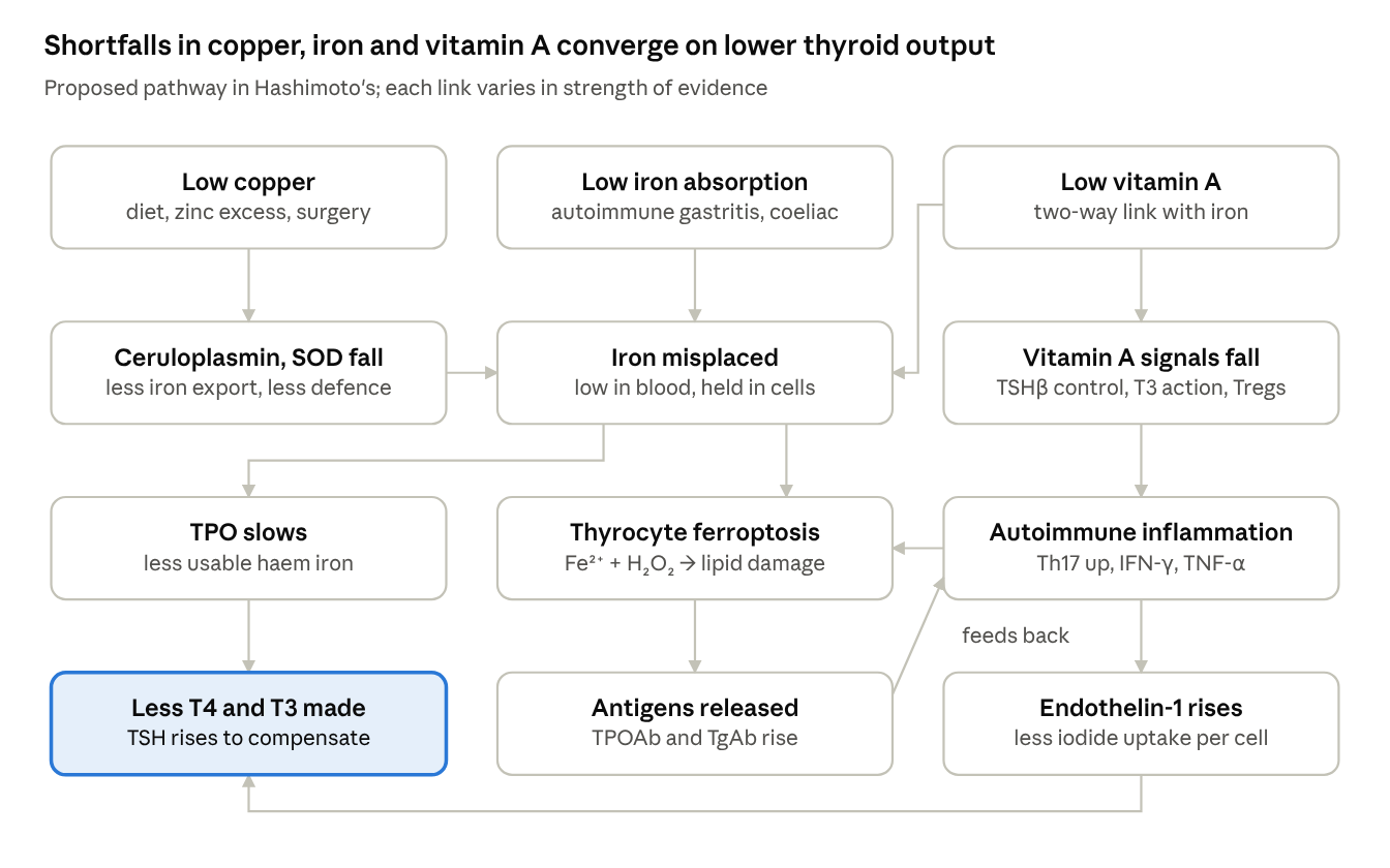
Read top to bottom: the left column ends in lower hormone output, the middle in antibody production, and the right column loops back into iron damage. Several links (copper → iron trapping, vitamin A → iron, ferroptosis in Hashimoto’s) are supported by animal, genetic or tissue studies rather than trials in thyroid patients.
Expected effect on thyroid hormones and antibodies
| Factor | TSH | T4/T3 output | Antibodies | Evidence strength |
|---|---|---|---|---|
| Low copper | No direct effect shown | Indirect, via iron handling and antioxidant loss | Mixed: higher, lower and no difference all reported | Weak; strongly confounded because thyroid status itself shifts copper |
| Iron deficiency | Mixed across studies | Lower free T4 and T3 (meta-analysis) | More TPOAb and TgAb positivity | Moderate: meta-analysis of observational studies plus animal TPO data |
| Iron trapped in thyrocytes | Rises as output falls | Lower, through cell loss | Higher, through antigen release | Early: cell and tissue studies |
| Systemic iron overload | Rises in transfusional overload | Lower in thalassaemia; usually preserved in haemochromatosis | Not a main driver | Moderate, condition-specific |
| Low vitamin A | Higher (weaker TSHβ suppression) | Can stay normal or high with central resistance | Lower vitamin A linked to higher TgAb | Moderate for TSH (two RCTs); weak for autoimmunity |
| High endothelin-1 | No correlation | Lower iodide uptake in cell studies | Tracks autoimmune inflammation | Case-control plus cell studies |
Graves’ disease runs partly in reverse. High thyroid hormone raises serum copper, ceruloplasmin and ET-1, and these fall again with antithyroid treatment. In Graves’, these markers are more a consequence of hyperthyroidism than an upstream cause, although ET-1 may still contribute to gland growth and blood flow.
Testing, interpretation and safety
None of these four factors is checked in a routine thyroid work-up, and each test is distorted by inflammation or by thyroid status itself. Interpreting them together, and re-testing once euthyroid, gives a much fairer picture than any single result.
| What to ask about | Tests | Main interpretation trap |
|---|---|---|
| Thyroid status and autoimmunity | TSH, free T4, free T3, TPOAb, TgAb (TRAb if Graves’ suspected) | Copper, ceruloplasmin, hepcidin and ET-1 all shift with hormone levels, so read them alongside TFTs |
| Iron | Full blood count, ferritin, transferrin saturation, serum iron, CRP | Ferritin rises with inflammation; a "normal" ferritin with low transferrin saturation can mean iron is locked in storage |
| Copper | Serum copper, ceruloplasmin, serum zinc | Raised by oestrogen (pill, HRT, pregnancy) and inflammation; lowered by untreated hypothyroidism. A normal or high result does not show tissue copper is adequate; ask about ceruloplasmin oxidase activity where available |
| Vitamin A | Serum retinol (ideally with CRP) | Retinol falls during inflammation and with iron deficiency; it stays normal until liver stores are well depleted |
| Endothelin-1 | Research use only | Not a clinical test; not useful for staging disease |
Who might reasonably ask for copper testing: people with anaemia with low blood iron but normal or high ferritin, low neutrophils, unexplained neuropathy or balance problems, previous bariatric or bowel surgery, coeliac disease, or regular zinc supplements or zinc-containing denture fixative.
Safety before supplementing
- Zinc depletes copper at high doses; intakes above about 50 mg/day block copper absorption, and long-term zinc supplements without copper are a recognised route to deficiency.
- Copper: shortfall is far more common than excess. Many adults eat less than UK and EU intake targets, and high zinc intake drains copper further (see the copper section). Copper toxicity is rare and mainly seen in Wilson’s disease, a rare inherited disorder in which the liver cannot excrete copper — people with Wilson’s disease should not add copper. Food sources include shellfish, liver, nuts, seeds, cocoa and wholegrains.
- Vitamin A (preformed) toxicity comes overwhelmingly from supplements, not from food; this post does not recommend vitamin A supplements, and food sources such as liver, egg yolks, butter and oily fish are the preferred route (see the vitamin A section).
- Selenium powers GPX4, the enzyme that blocks ferroptosis, and the enzymes that protect thyroid cells from the hydrogen peroxide they make. This post does not recommend selenium supplements; food sources include Brazil nuts, fish and shellfish, eggs and offal.
Checking the claims in the starting notes
| Claim in the notes | Verdict |
|---|---|
| Copper deficiency is rare | Overstated. Frank deficiency is uncommon; under-diagnosis and low intake are common (see copper section) |
| Ferroptosis and the Thyroid, Metabolites, Nov 2026 | Verified — published as Metabolites 16(11):850 |
| ET-1 study shows rise driven by MCP-1 and MIP-1α | Partly. The 2003 paper attributes raised ET-1 to autoimmune inflammation; the chemokine mechanism comes from earlier lab work it cites, not from measurements in these patients |
| Retinoic acid supports ceruloplasmin production | Supported in rats (Barber & Cousins, J Nutr 1987); not shown in humans. Vitamin A-deficient rats actually had raised ceruloplasmin, attributed to inflammation |
| Iron overload causes hypothyroidism | True for transfusional overload (thalassaemia); weak for hereditary haemochromatosis, where thyroid function is usually preserved |
| Copper deficiency lowers TRH activity | No direct human evidence found; treat as hypothesis |
| ET-1 drives hypervascularity in Graves' | Plausible — ET-1 is mitogenic for thyroid cells — but VEGF and TSH-receptor stimulation are the better-established drivers |
| Hashimoto's patients often have low iron | Correct, and important: deficiency and cellular trapping are not mutually exclusive |
This article is educational and not a substitute for individual clinical advice. Anyone with thyroid disease should discuss testing and supplements with their GP or endocrinologist.
The same four factors across other autoimmune diseases: Autoimmune disease beyond the thyroid
Sources
Copper status and deficiency
- NIH Office of Dietary Supplements — Copper fact sheet for health professionals
- Altarelli et al. Copper deficiency: causes, manifestations, and treatment. Nutr Clin Pract 2019
- Myint et al. Copper deficiency anemia: review article. Ann Hematol 2018
- Halfdanarson et al. Hematological manifestations of copper deficiency. Eur J Haematol 2008
- Copper deficiency: like a bad penny. Practical Gastroenterology 2020
- Review on copper and zinc intake and RDAs (PMC8970836)
- Derbyshire. Micronutrient intakes of British adults across mid-life (NDNS). Front Nutr 2018
- Nordic Nutrition Recommendations 2023 — copper scoping review
- Hypocupraemia secondary to zinc-containing dental adhesive. BMJ Case Rep 2021
- UK Committee on Toxicity — copper discussion paper
Copper and the thyroid
- Bhat et al. Copper and ceruloplasmin in hypothyroidism and Graves’. MRIMS J Health Sci 2024
- Hashimoto’s thyroiditis and hypoceruloplasminaemia. ESPE 2015 abstract
- Szczepanik et al. Zinc, copper and antioxidant status in women with Hashimoto’s. IJERPH 2021
- Trace elements and thyroid antibodies in newly diagnosed Hashimoto’s. Int J Endocrinol Metab 2024
- Qiu et al. Micronutrients and autoimmune thyroiditis: Mendelian randomisation. Front Immunol 2024
- Copper, iron, zinc and selenium in Hashimoto’s. Sakarya Med J 2018
- Thyroid hormones in copper-deficient rats. Z Ernährungswiss 1996
Iron
- Garofalo et al. Iron deficiency and thyroid function: systematic review and meta-analysis. Nutrients 2023
- Gierach et al. Iron and ferritin deficiency in women with hypothyroidism and Hashimoto’s. Endokrynol Pol 2024
- Rayman. Multiple nutritional factors and thyroid disease. Proc Nutr Soc 2019
- Hernik et al. Hepcidin falls after restoring euthyroidism in Hashimoto’s. Sci Rep 2019
- GeneReviews — Aceruloplasminemia
- Ceruloplasmin and iron — IntechOpen chapter
- Thyroid dysfunction and iron overload in HbE/β-thalassaemia. Arch Med Health Sci 2019
- Thyroid function in genetic haemochromatosis. BES 2002 abstract
- TSH and free T4 in the HEIRS haemochromatosis screening study
- Ferroptosis in Hashimoto's thyroiditis. Endocrine Abstracts vol 110
- Ferroptosis and the Thyroid. Metabolites 2026;16(11):850
- Zimmermann et al. Vitamin A mobilises iron stores and raises haemoglobin without changing body iron. Am J Clin Nutr 2006
- Bloem. Interdependence of vitamin A and iron. Proc Nutr Soc 1995
- Iron studies in absolute vs functional iron deficiency (PMC10485845)
Vitamin A
- Zimmermann et al. Vitamin A deficiency and supplementation in goitrous children. JCEM 2004
- Zimmermann. Interactions of vitamin A and iodine deficiencies on the pituitary-thyroid axis
- Farhangi et al. Vitamin A supplementation and thyroid function in premenopausal women. J Am Coll Nutr 2012
- Elias et al. Retinoic acid inhibits Th17 polarisation and enhances FoxP3. Blood 2008
- Retinoic acid increases Foxp3+ Tregs and inhibits Th17 via Smad3. J Immunol
- Raverdeau & Mills. Modulation of T cell responses by retinoic acid. J Immunol 2014
- Liang et al. Vitamin A and E status across Hashimoto's in women. Front Nutr 2026
- Mejia & Erdman. Vitamin A deficiency, iron metabolism and anaemia. Nutr Rev 2025
- Li et al. Iron repletion restores hepatic vitamin A mobilisation in rats. 2020
- Barber & Cousins. Induction of ceruloplasmin by retinoic acid in rats. J Nutr 1987
- Hodges et al. Hematopoietic studies in vitamin A deficiency. Am J Clin Nutr 1978
Endothelin-1
- Endothelin-1 levels in patients with disorders of the thyroid gland. Exp Clin Endocrinol Diabetes 2003
- Letizia et al. Increased plasma ET-1 in hyperthyroidism. Metabolism 1995
- Chu et al. Hyperthyroidism and plasma ET-1. Exp Biol Med 2006
- Mayyas et al. ET-1 and atrial fibrillation in hyperthyroidism. PLoS One 2018
- ET-1 and porcine thyroid cells: inhibition of iodide uptake. J Endocrinol 1994
- Tseng et al. Endothelin production by human thyroid follicular cells
- Colin et al. ET-1 gene expression in rat thyroid during goitre. J Endocrinol 1994